Urine-based early-stage bladder cancer examination method and system

An inspection method and bladder cancer technology, applied in the field of urine-based early bladder cancer inspection and system, can solve the problems of "impossible inspection, easy missed inspections, and scarcity of expert doctor resources."

Inactive Publication Date: 2014-05-07
蔡志明 +1
View PDF5 Cites 1 Cited by
  • Summary
  • Abstract
  • Description
  • Claims
  • Application Information

AI Technical Summary

Problems solved by technology

[0013] 1) The formation process is seldom discovered. If hematuria and other discomfort signs are found to be checked again, it is often already in the middle and late stage
[0014] 2) After being magnified thousands of times under the microscope, a standard pathological slide needs about tens of thousands to millions of "fields of view". At present, the resources of expert doctors are scarce, and most of them are done in the case of "suspect" by outpatient doctors. Final diagnosis, impossible to achieve "check"
[0015] 3) The inspection process is extremely painful. The current inspection method is mostly cystoscopy. For example, males need to insert from the front of the genitals to the bladder.
[0016] 4) It is very easy to miss inspection - the bladder is an "inverted pear shape", the effective inspection area of ​​cystoscopy is only a few square centimeters, and the inner wall surface area is about thousands of square centimeters

Method used


Image

  • Urine-based early-stage bladder cancer examination method and system
  • Urine-based early-stage bladder cancer examination method and system
  • Urine-based early-stage bladder cancer examination method and system

Examples


Embodiment Construction

[0037] The present invention will be further described in detail below through specific embodiments in conjunction with the accompanying drawings.

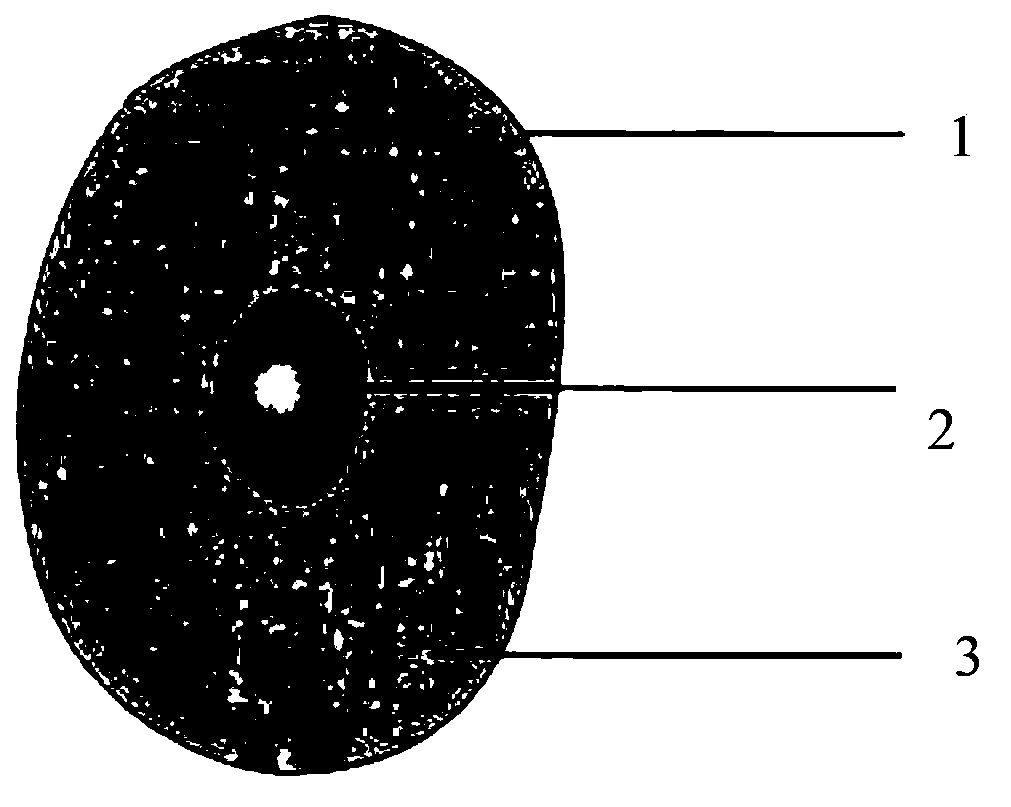
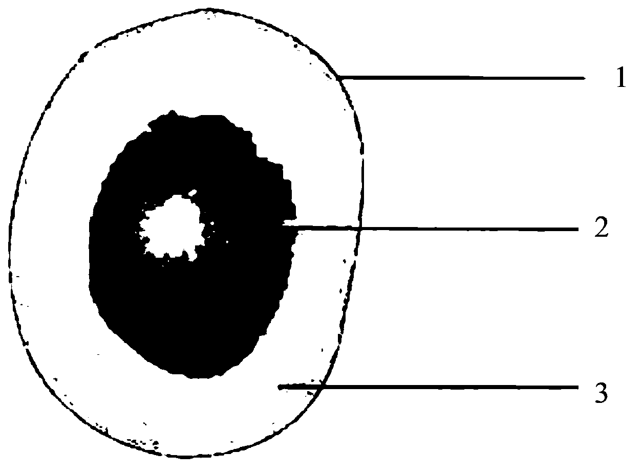
[0038] Such as figure 1 As shown, the cell is composed of three parts: cell membrane 1 (cell membrane), cytoplasm 2 (cytoplasm) and nucleus 3 (nucleus). Such as figure 2 As shown, for the diseased cancerous cells, the obvious feature is that the nucleus expands and the cytoplasm decreases, that is, the area of ​​the nucleus increases and the area of ​​the cytoplasm decreases.

[0039] Let S1 be the nucleus area, S2 be the cytoplasm area, K=S1 / S2.

[0040] For normal cells, k

[0041] For early cancer cells, a

[0042] For metaphase cancer cells, b

[0043] For advanced cancer cells, c

[0044] The above-mentioned range of the K value may be a range of empirical values ​​obtained through statistics after a limited number of experiments and data collection.

[0045] Early detection of bladder cancer can be ...


PUM


Abstract

The invention discloses a urine-based early-stage bladder cancer examination method and system. The method comprises the following steps: (a) making a pathology slide of urine; (b) acquiring an image of the pathology slide through an image acquisition mechanism with a microscopic lens; (c) acquiring the area ratio K of cell nucleuses and cytoplasms of all cells in the image; (d) comparing the area ratio K and a preset area ratio range, and examining suspected cancerous cells according to the comparison result. With adoption of the method and system, complete examination and comparison of the acquired cell information can be realized so as to rapidly acquire the examination result of the cancerous cells, as a result, accurate first-hand data is provided for a doctor to confirm cancerous cells in a follow-up process.

Description

technical field [0001] The invention relates to a urine-based early bladder cancer detection method and system. Background technique [0002] Cancer is one of the most common fatal diseases in the world today. Millions of people die from cancer every year around the world. In the process of facing this century-old disease of cancer, human beings have never found effective treatments and drugs. [0003] The proliferation speed of cancer cells is calculated by doubling time, 1 becomes 2, 2 becomes 4...and so on. The average doubling time of gastric cancer, intestinal cancer, liver cancer, pancreatic cancer, and esophageal cancer is 31 days; the doubling time of breast cancer is more than 40 days. As cancer cells multiply, the more advanced the cancer, the faster it develops. [0004] Cancer can be divided into 3 stages, [0005] The first third can be prevented and cured; [0006] One-third of the symptoms can be alleviated through treatment, early detection and early trea...

Claims


Application Information

Patent Timeline
Patent Type & Authority Applications(China)
IPC IPC(8): G01N21/85
Inventor 董涛蔡志明孙小娟吴松关弘
Owner 蔡志明
Why Eureka
Try Eureka