Separated imaging method for superimposed targets based on multi-spectrum X rays

A target separation and imaging method technology, applied in the field of medical imaging, can solve the problems of low atomic number material occlusion, high contrast of X-ray images, low image contrast, etc., to improve observation effect, facilitate judgment, and overcome the shortcomings of image overlap

Inactive Publication Date: 2014-05-28
NANJING UNIV OF POSTS & TELECOMM
View PDF3 Cites 14 Cited by
  • Summary
  • Abstract
  • Description
  • Claims
  • Application Information

AI Technical Summary

Problems solved by technology

The X-ray attenuation of high atomic number materials is significantly greater than that of low atomic number materials, resulting in high contrast of X-ray images of high atomic number materials and low image contrast of low atomic number objects
Therefore, low atomic number materials are easily obscured by materials superimposed on top of them in an X-ray image

Method used


Image

  • Separated imaging method for superimposed targets based on multi-spectrum X rays
  • Separated imaging method for superimposed targets based on multi-spectrum X rays

Examples


Embodiment Construction

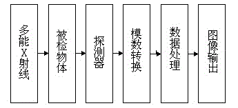
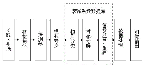
[0016] According to the multi-energy spectrum X-ray-based superposition target separation imaging method of the present invention, the imaging equipment is an X-ray source, an imaging object, an X-ray detector, a CCD, a data analysis and processor, and a terminal output device in order of placement; first Take multiple X-ray images in the same field of view by changing the X-ray source voltage; combine the X-ray attenuation imaging formula and image grayscale to describe the imaging process of multiple X-ray images to establish the X-ray attenuation imaging equation Solving the X-ray attenuation imaging equations by using independent component analysis technology to obtain the attenuation thickness of the superimposed target; reconstructing the three-dimensional image of each superimposed target according to the thickness of the superimposed target is to separate and image the superimposed target in the original imaging object.

[0017] The concrete steps of the embodiment of t...


PUM


Abstract

The invention provides a separated imaging method for superimposed targets based on multi-spectrum X rays. Imaging equipment sequentially comprises an X ray source, an imaging object, an X ray detector, a CCD (Charge Coupled Device), a data analyzing and processing device and terminal output equipment, which are put by an order. First, multiple X ray images are respectively shot for the imaging object in the same field of view through the change of the voltage of the X ray source. The imaging processes of the multiple X ray images are respectively described in combination of an X ray attenuation imaging formula and the gradation of the image so as to establish an X ray attenuation imaging equation set. The X ray attenuation imaging equation set is solved by an independent component analysis technology so as to obtain the attenuation thickness of the superimposed targets. The three-dimensional image of each superimposed target is reconstructed according to the thickness of the superimposed targets, that is, the superimposed targets in the original imaging object are separately imaged. Compared with a conventional X ray imaging method, the method has the advantages that the visual effect of the images is improved, and the medical diagnosis accuracy is improved.

Description

technical field [0001] The invention relates to diagnosis and detection by using X-ray imaging in the fields of medical imaging and non-destructive testing, and specifically relates to a new technology of X-ray stereoscopic imaging. Background technique [0002] X-rays are widely used in medical diagnosis, industrial flaw detection and non-destructive testing. A typical X-ray imaging system is mainly composed of an X-ray source, an X-ray detector, a data processing unit, and an image output device. An X-ray image is formed by recording the attenuated X-ray intensity that penetrates an object. The material attenuation coefficient varies with the ray energy and is determined by the chemical composition of the material. The X-ray attenuation of high atomic number materials is significantly greater than that of low atomic number materials, resulting in high contrast of X-ray images of high atomic number materials and low image contrast of low atomic number objects. Therefore, ...

Claims


Application Information

Patent Timeline
Patent Type & Authority Applications(China)
IPC IPC(8): G01N23/04G01N23/083
Inventor 喻春雨
Owner NANJING UNIV OF POSTS & TELECOMM
Why Eureka
Try Eureka