Blood vessel ROI dividing method based on intravascular ultrasonic image

A technology for ultrasound images and blood vessels, which is applied in image analysis, image data processing, ultrasound/sonic/infrasonic diagnosis, etc. It can solve the problem that the initial contour is not easy to determine, the diagnosis result cannot reflect the actual situation very objectively, and the accuracy of statistical modeling is reduced. And other issues

Inactive Publication Date: 2014-06-25
BEIJING UNIV OF TECH
View PDF2 Cites 24 Cited by
  • Summary
  • Abstract
  • Description
  • Claims
  • Application Information

AI Technical Summary

Problems solved by technology

In the region of interest of the IVUS image, the doctor often needs to visually or empirically determine the edge of the adventitia in the luminal membrane and the outline of the plaque, which inevitably leads to a diagnosis that cannot reflect the actual situation very objectively, and also gives the doctor bring some work difficulties
[0004] Therefore, it is necessary to quickly, accurately and automatically segment intravascular ultrasound images by computer. At present, there are three main computer segmentation algorithms for intravascular ultrasound images: the first is the image segmentation method based on statistics, document 1 ( G. Mendizabal-Ruiz, M. Rivera, et al., "A probabilistic segmentation method for the identification of luminal borders in intravascular ultrasound images", IEEE Conference on Computer Vision and pattern Recognition, pp.1-8, 2008.) via Statistical modeling is performed on the gray distribution of IVUS images to achieve the segmentation of intravascular ultrasound images, but the existence of more complex image features such as artifacts and plaques in intravascular ultrasound images will greatly reduce the accuracy of statistical modeling
The second method is mainly to achieve intravascular ultrasound image segmentation by means of machine learning, but the model of this method is too complicated, and it is often subject to more restrictions in actual application. The third method is an algorithm based on the active contour model. Document 2 (Gong Yanxin, Sun Fengrong, etc., "Edge Extraction of Intravascular Ultrasound Images Based on Blood Speckle Noise Suppression and T-Snake Model", Chinese Journal of Image and Graphics, Vol.12, No.4, pp.655-660, 2007) Although the proposed method can obtain good segmentation results, it depends on the selection of the initial contour line, especially for IVUS images with blurred edges and high texture self-similarity, the initial contour is more difficult to determine, which also affects to a certain extent. The accuracy of this segmentation method

Method used


Image

  • Blood vessel ROI dividing method based on intravascular ultrasonic image
  • Blood vessel ROI dividing method based on intravascular ultrasonic image
  • Blood vessel ROI dividing method based on intravascular ultrasonic image

Examples


Embodiment Construction

[0091] The present invention is realized by adopting the following technical means:

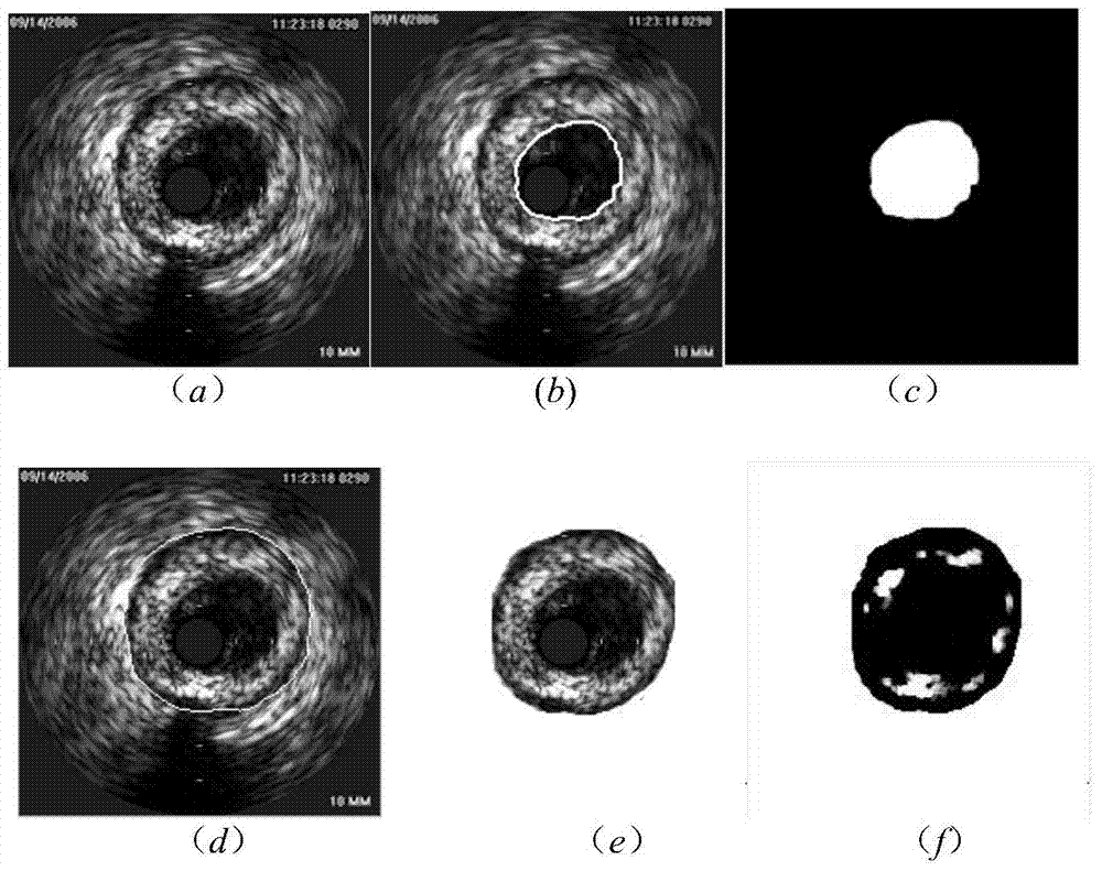
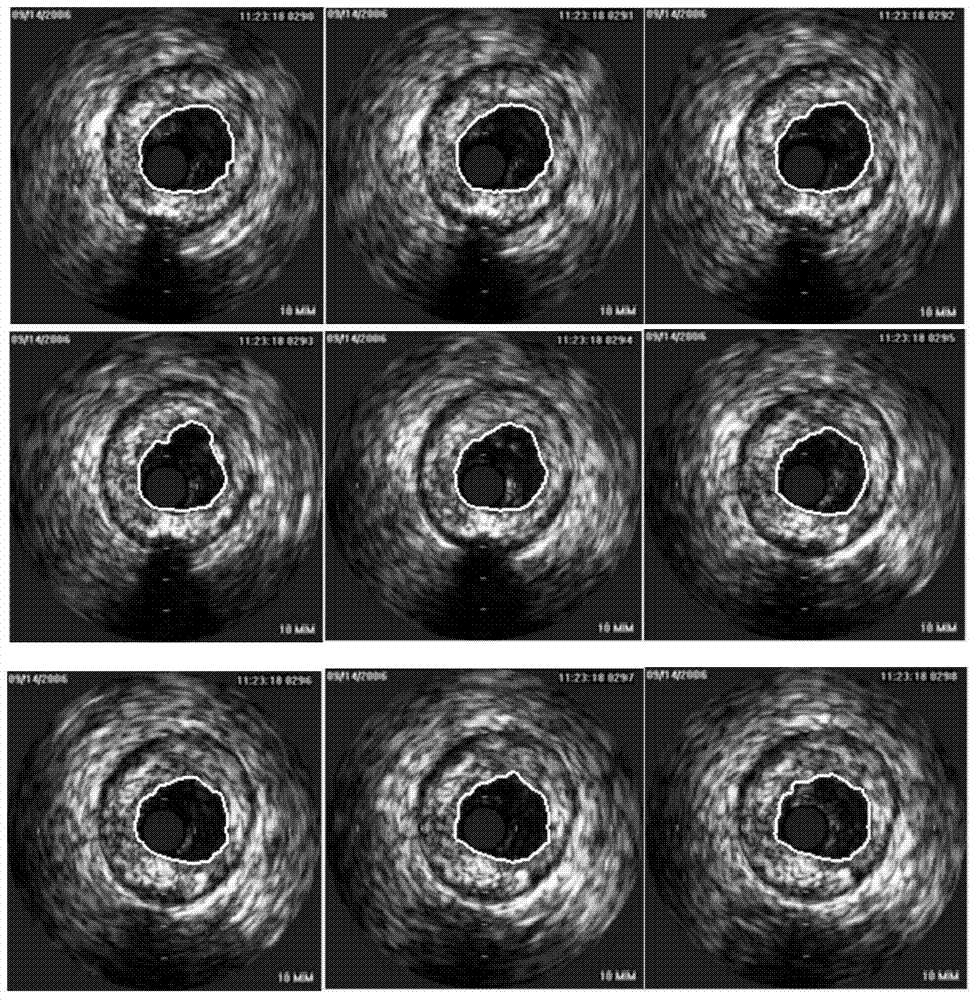
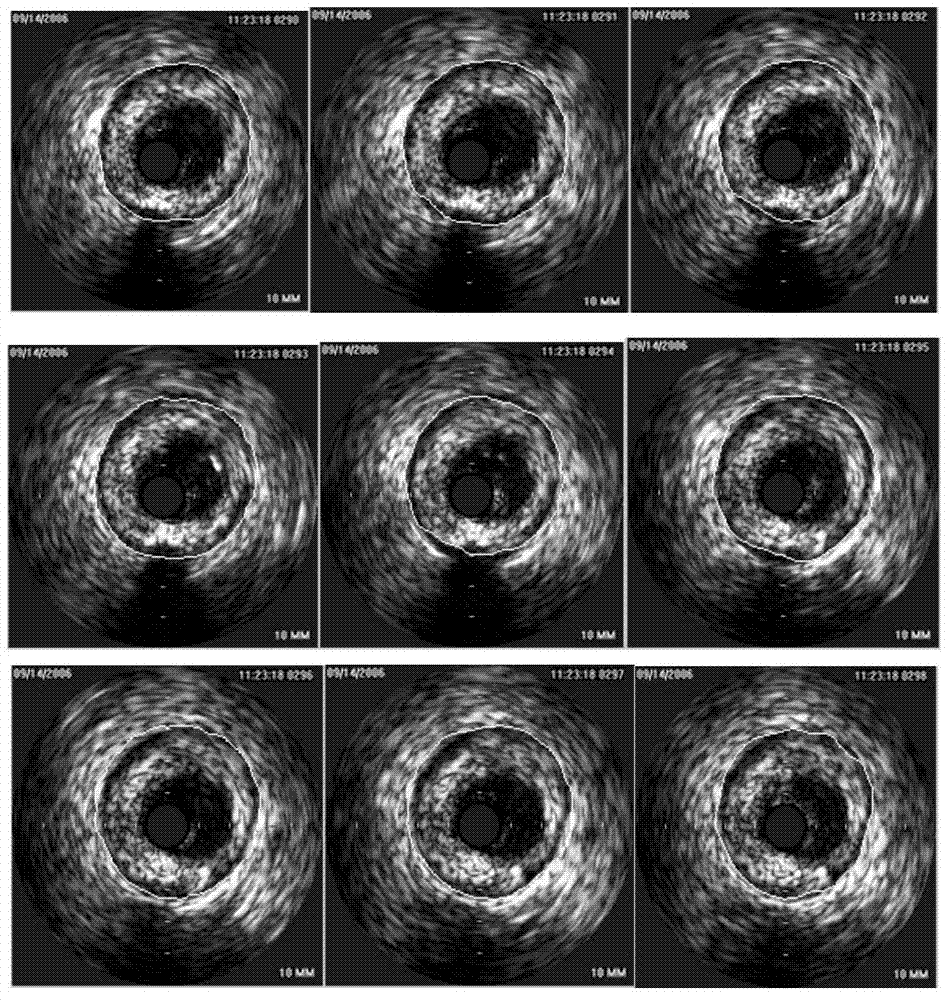
[0092] A vascular ROI segmentation method based on intravascular ultrasound images. Firstly, the image to be segmented is combined with the improved level set model algorithm and the narrow-band method to realize the segmentation of the luminal membrane in the intravascular ultrasound image. Then, the center of the luminal area is located and the initial contour curve of the Snake model is generated for iterative convergence to obtain the inner and outer parts of the vessel. The edge of the membrane, and finally, select the region inside the media-adventitia as the ROI of the plaque, and combine the algorithm of the global minimum active contour model to realize the segmentation of the plaque contour of the blood vessel ROI.

[0093] The above-mentioned blood vessel ROI segmentation method based on the intravascular ultrasound image comprises the following steps:

[0094] Step 1: Using an in...


PUM


Abstract

The invention relates to a blood vessel ROI dividing method based on an intravascular ultrasonic image. The method includes the steps that a lumen area and a lumen membrane outline of a blood vessel are divided firstly, the initial outline of a parameter active outline model is obtained by positioning the center of the lumen area, then the middle and outer membrane outline curve of the blood vessel is obtained through convergence, and priori knowledge of the lumen area information is used sufficiently for extracting a middle and outer membrane; an inner area is taken as ROI for the middle and outer membrane outline curve, and blood vessel plaques are divided by minimizing a movable outline model globally. Visualization of outline information of the middle and outer membrane and the plaques of the lumen membrane of the blood vessel ROI is achieved, in comparison with an IVUS image dividing method based on statistics, a complex statistical modeling process of the IVUS image dividing method is abandoned, and the dividing result is not affected by IVUS image artifacts and plaque features; the step of dividing the initial outline of the edge of the middle and outer membrane in IVUS images in advance is omitted, and dividing efficiency is improved.

Description

technical field [0001] The invention relates to the field of computerized medical image analysis, in particular to a method for segmenting a region of interest (ROI) of a blood vessel based on an IVUS (Intravascular Ultrasound) image. Background technique [0002] At present, in most countries, cardiovascular and cerebrovascular diseases have gradually become one of the most important factors of human death. Coronary atherosclerotic lesions are the main cause of myocardial infarction and cerebral infarction. If early recognition and diagnosis of part of the sclerotic lesion can be achieved, it will be of great significance to the diagnosis and treatment of coronary artery disease. IVUS is such an ultrasound diagnostic method for cardiovascular diseases. Intravascular ultrasound images can display real-time plaque morphology of vessel walls for doctors, thereby providing guidance for the clinical diagnosis of cardiovascular diseases. [0003] However, for the collected IVUS ...

Claims


Application Information

Patent Timeline
Patent Type & Authority Applications(China)
IPC IPC(8): G06T7/00A61B8/00
Inventor 汪友生王志东李冠宇
Owner BEIJING UNIV OF TECH
Why Eureka
Try Eureka