CT (computed tomography) imaging method and CT imaging system based on multi-mode Scout scanning

A technology of CT imaging and scanning table, which is applied in computerized tomography scanners, instruments for radiological diagnosis, medical science, etc. It can solve the problems that it cannot be used as coronary artery screening, high radiation dose, low contrast and material overlap, etc. Achieve the effects of shortening imaging time, reducing X-ray dose, and lower X-ray dose

Inactive Publication Date: 2014-07-02
GE MEDICAL SYST GLOBAL TECH CO LLC
View PDF11 Cites 24 Cited by
  • Summary
  • Abstract
  • Description
  • Claims
  • Application Information

AI Technical Summary

Problems solved by technology

However, compared with ordinary Scout scanning, the radiation dose of various spiral CTs including 64-slice spiral CT and GSI 64-slice spiral CT is still higher, and the images acquired by ordinary Scout scanning are due to the existence of lower contrast and material overlap. not as an image for coronary artery screening

Method used


Image

  • CT (computed tomography) imaging method and CT imaging system based on multi-mode Scout scanning
  • CT (computed tomography) imaging method and CT imaging system based on multi-mode Scout scanning
  • CT (computed tomography) imaging method and CT imaging system based on multi-mode Scout scanning

Examples


Embodiment Construction

[0049] In the following detailed description, some exemplary embodiments according to the present invention are described with reference to the accompanying drawings. Those skilled in the art will appreciate that the present invention is not limited to these exemplary embodiments.

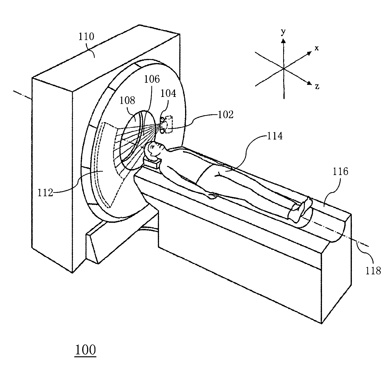
[0050] Figures 1A-1B A radiation CT system 100 according to an exemplary embodiment of the present invention is shown. In one embodiment, the radiation CT system 100 is an X-ray CT system.

[0051] Such as Figures 1A-1B As shown, the X-ray CT system 100 mainly includes three parts: a scanning gantry 110 , a scanning support table 116 supporting and positioning an examinee 114 , and an operation console 130 . Scanning gantry 110 includes X-ray tube 102 . The X-ray 106 radiated from the X-ray tube 102 is shaped by the collimator 104 to obtain an X-ray beam such as a fan beam or a cone beam, and is irradiated onto the region of interest of the subject 114, and is sensed from the subject 114. The ...


PUM


Abstract

A CT imaging method and a CT system based on a multi-mode scout scan. The CT imaging method based on a multi-mode scout scan comprises: performing an instant switching dual energy scout radiation scan on a region of interest of a subject by way of instant switching between high voltage and low voltage to collect dual energy protection data of the region of interest; and reconstructing a material decomposition image and a mono-energetic image based on the collected dual energy projection data.

Description

technical field [0001] The present application relates to the field of radiation CT, and more specifically, relates to a CT imaging method and a CT system based on multi-mode Scout (positioning) scanning. Background technique [0002] At present, radiation CT systems such as X-ray CT systems are widely used in various medical institutions to perform three-dimensional imaging of regions of interest of subjects, such as coronary arteries of subjects, to help clinicians The examiner makes an accurate medical diagnosis. [0003] Among the existing coronary artery screening methods, intracoronary ultrasound area measurement is one of the methods to accurately determine the degree of coronary artery stenosis, but this method has high requirements for technical conditions and high cost of funds, so it is not suitable for routine clinical application . [0004] DSA (Digital Subtraction Angiograph, Digital Subtraction Angiography) Coronary angiography, the catheter is inserted thro...

Claims


Application Information

Patent Timeline
Patent Type & Authority Applications(China)
IPC IPC(8): A61B6/03
CPCA61B6/481A61B6/032A61B6/482A61B6/027A61B6/488A61B6/504
Inventor 李颖赵静陈雨丝魏东
Owner GE MEDICAL SYST GLOBAL TECH CO LLC
Why Eureka
Try Eureka