SPECT (single-photon emission computed tomography) imaging method based on ordered subset algorithm

An imaging method and subset algorithm technology, applied in the field of biomedical imaging, can solve the problem of not fully highlighting the speed advantage of the OS-EM algorithm, and achieve the effects of reducing photon counts, reducing demand, and shortening acquisition time.

Active Publication Date: 2014-09-10
PEKING UNIV
View PDF4 Cites 5 Cited by
  • Summary
  • Abstract
  • Description
  • Claims
  • Application Information

AI Technical Summary

Problems solved by technology

Considering that the OS-EM algorithm has the highest efficiency in the first few iterations, the ind

Method used


Image

  • SPECT (single-photon emission computed tomography) imaging method based on ordered subset algorithm
  • SPECT (single-photon emission computed tomography) imaging method based on ordered subset algorithm
  • SPECT (single-photon emission computed tomography) imaging method based on ordered subset algorithm

Examples


Embodiment Construction

[0028] The present invention will be further described through the embodiments below in conjunction with the accompanying drawings.

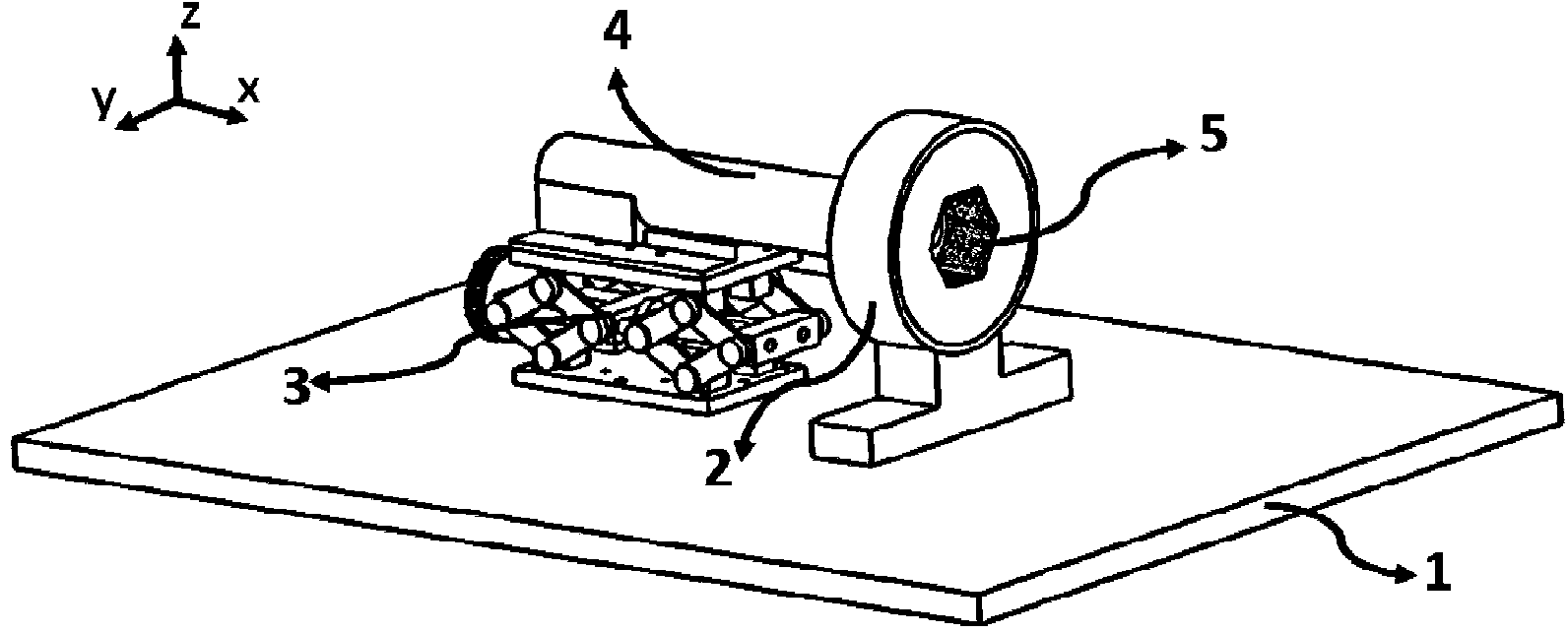
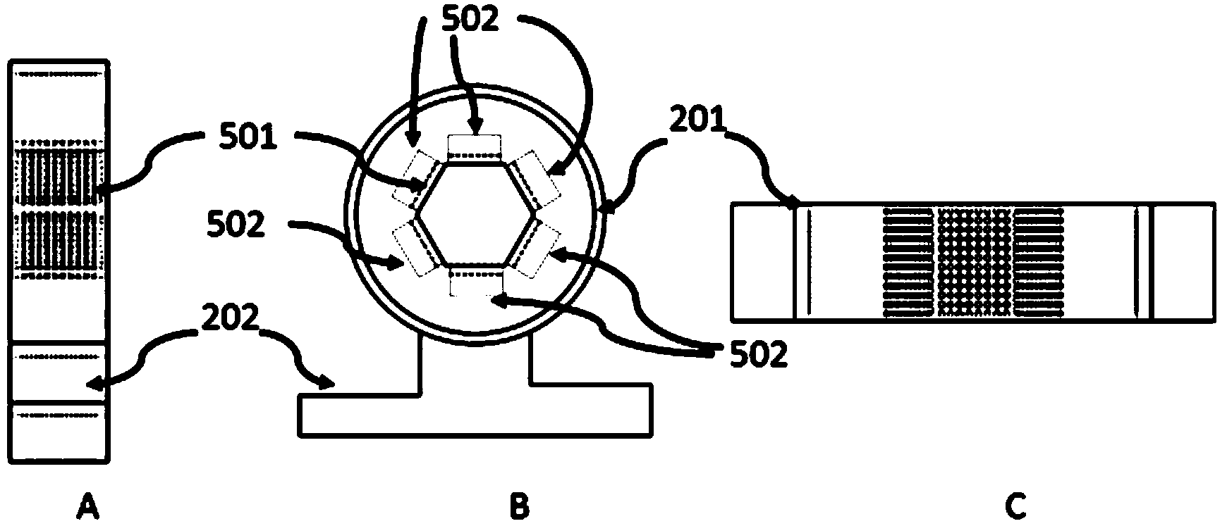
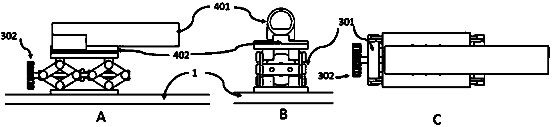
[0029] Such as figure 1 As shown, the present embodiment takes a hexagonal detector: M=6 as an example, and the SPECT imaging device of the ordered subset algorithm includes: a rotating frame 2, an elevating table 3, an examination bed 4, a SPECT detector and a collimator device 5 and a data acquisition system; wherein, the rotating frame 2 is fixed on one end of the base plate 1, and the rotating frame 2 has a through hole, which rotates with the axis of the through hole as the axis of rotation during imaging; the SPECT detector and the collimator 5 are arranged Around the through hole of the rotating frame, and arranged in an equilateral hexagon, the collimator is located between the SPECT detector and the imaging area; the lifting table 3 is fixed on the other end of the bottom plate 1; the examination bed 4 is fixed on the lifting table 3 A...


PUM


Abstract

The invention discloses a SPECT (single-photon emission computed tomography) imaging method based on an ordered subset algorithm. The SPECT imaging method includes that a SPECT detector and a collimator are arranged in an equilateral polygon arranged on the periphery of a through hole of a rotating frame, the number of edges of the polygon corresponds to the number of angles in one subset, the rotating frame is rotated to have data collected, each time of data collection is accompanied with OS-EM iteration once, and after multiple iterations, images meeting requirements of image resolution are reestablished through a computer. The SPECT detector and the collimator are in polygon arrangement matched with the ordered subset algorithm, the advantages of the algorithm can be effectively exerted by means of hardware grouping, collecting full 360 angles is not needed, unnecessary photon counting is effectively reduced, and collecting time is shortened; besides, quantity demanded on the detectors and the collimators is decreased by adopting the rotating frame, and economical efficiency is high.

Description

technical field [0001] The invention relates to the field of biomedical imaging, in particular to a SPECT imaging method based on an ordered subset algorithm. Background technique [0002] Single-Photon Emission Computed Tomography (SPECT) uses radionuclide tracer technology to obtain projection data by detecting the rays emitted by radioisotope drugs ingested by certain organs in the organism, and then obtain the projection data. The projection data is reconstructed into a tomographic image, and then the distribution and metabolism information of specific molecules in the object to be measured can be obtained. Small animal SPECT imaging can accurately reflect the dynamic process of uptake and metabolism of radionuclide-labeled drugs in small animals, and monitor the intervention effect of treatment methods on diseases. In addition, since SPECT imaging can be used for continuous longitudinal studies on the same small animal model, errors caused by individual differences in ...

Claims


Application Information

Patent Timeline
IPC IPC(8): A61B6/03
Inventor 任秋实谢肇恒李素莹刘琪刘曦张秋实卢闫晔周坤田涧
Owner PEKING UNIV
Why Eureka
Try Eureka