Methods for diagnosis and/or prognosis of gynecological cancer
A technology for survival prognosis and ovarian cancer, applied in the fields of biochemical equipment and methods, microbial determination/examination, etc., which can solve the problems of undisclosed, information contradiction of the role of MECOM locus, etc.
- Summary
- Abstract
- Description
- Claims
- Application Information
AI Technical Summary
Problems solved by technology
Method used
Image
Examples
Embodiment 1
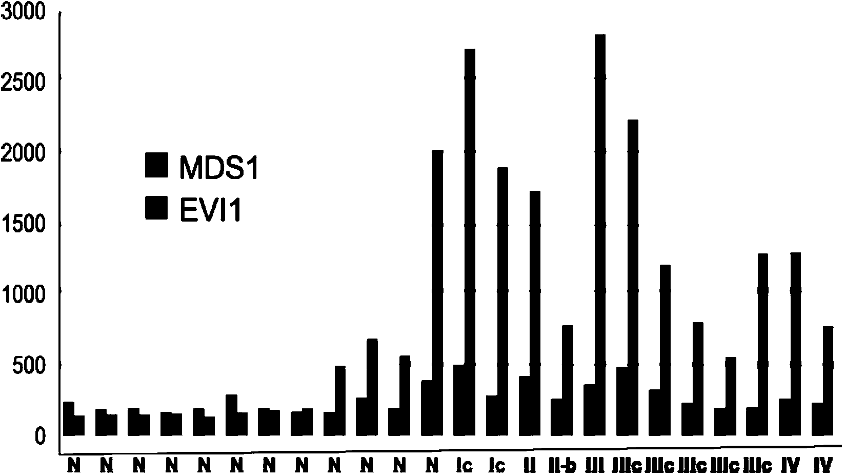
[0160] EVI1 gene expression is a distinguishing marker between normal and cancerous ovarian tissue
[0161] result in figure 1 , where expression of the EVI1 and MDS1 genes showing the MECOM locus is greatly increased in ovarian cancer cells relative to normal epithelial cells. In samples obtained from ovarian tissue using laser microdissection, the average expression of EVI1 and MDS1 genes was 3.6 and 1.4 times higher in cancerous tissues than in adjacent normal tissues ( figure 1 ). This difference in mean expression values was more significant for EVI1 relative to MDS1 (p=0.021). MDS1 and EVI1 expression values were associated with all samples (Pearson'sr=0.84, Kendall'sr=0.60).
Embodiment 2
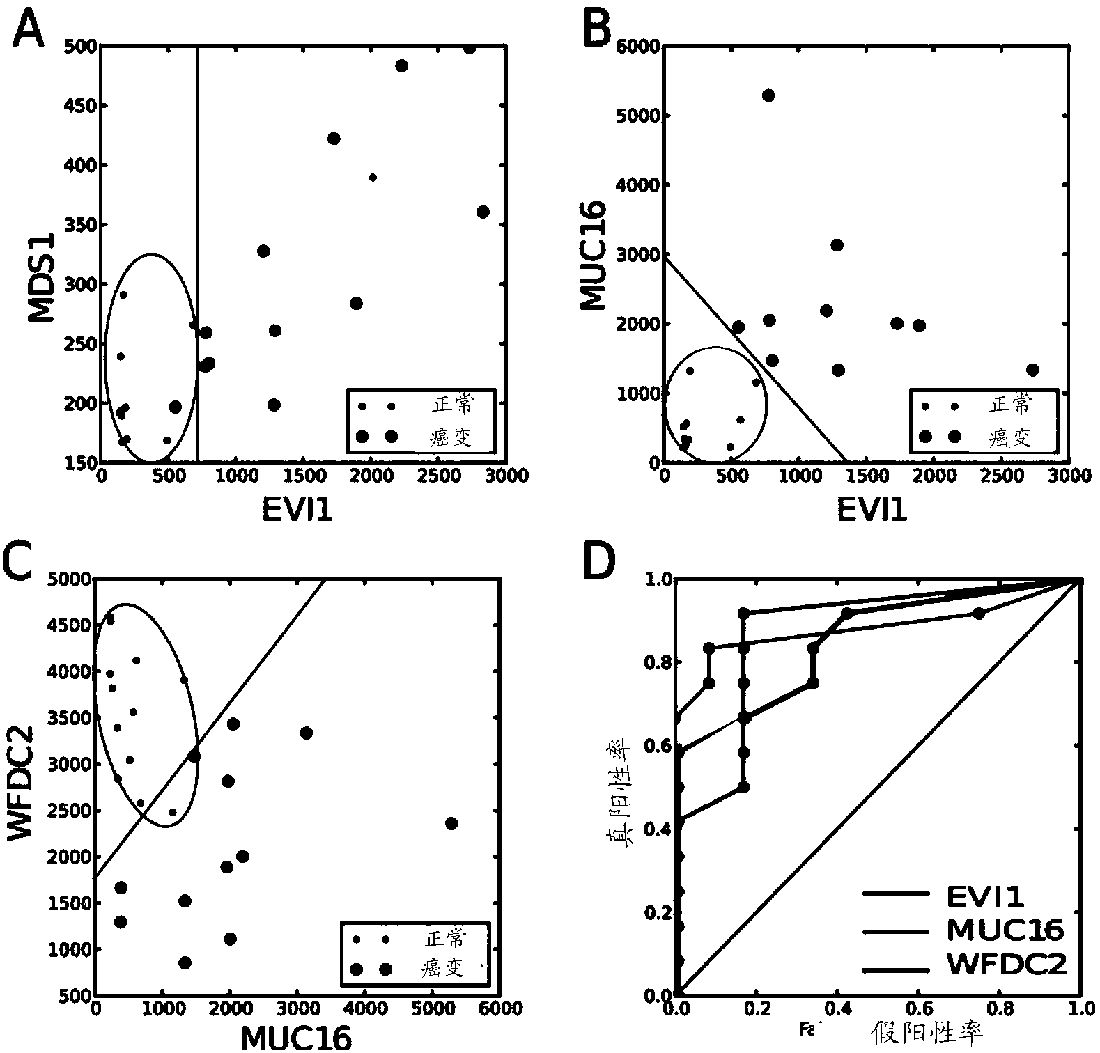
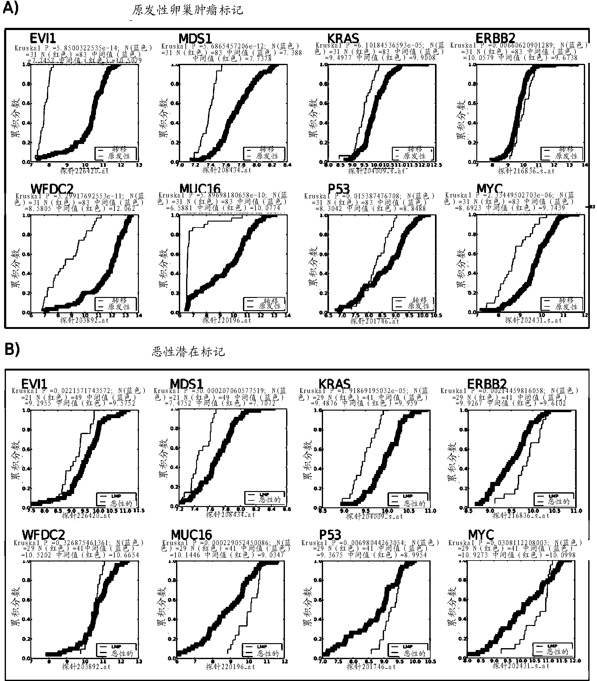
[0162] Example 2 EVI1 expression is a sensitive and specific marker of primary EOC tumors among traditional diagnostic biomarkers
[0163]To assess the importance of EV1 in ovarian cancer, its expression was analyzed in three datasets and its importance as a diagnostic marker in the gene set was re-examined, its importance in ovarian cancer diagnosis and patient survival is widely accepted : KRAS, ERBB2, P53, MYC, MUC16 (CA-125) and WFDC2 (HE4). Results for WFDC2 and MUC16 are shown in figure 2 middle. It can be known from the results that MECOM expression is different in normal ovarian epithelial cells and cancerous ovarian epithelial cells. Combinations of expression values of MECOM genes that differentiate EOC with false negative (FN)=1 and false positive (FP)=1 ( figure 2 (A)), which can be compared with figure 2 (C) Combination comparison of MUC16 (CA-125) and WWFDC2 (HE-4) markers. exist figure 2 (B) Perfect separation of the EV1 and MUC16 gene combinations w...
Embodiment 3
[0177] EVI1 expression is a sensitive marker of potential malignancy in EOC tumors
[0178] Regarding the potential malignancy of the tumor, the EVI1 gene ranked 5th (P=0.022) in terms of panel discrimination significance, surpassing the biomarkers MYC (P=0.03) and ERBB2 (P=0.74).
[0179] The specificity=21 / (21+4)=84% and the sensitivity=49 / (49+18)=73% of EVI1EVI1 at the expression cutoff level of 9.5, while the expression level of MUC16(CA-125) at the cutoff level of 9.7 Specificity=21 / (21+5)=81%, Sensitivity=49 / (49+11)=82%. exist Image 6 The comparison plot of the ROC curves shows that EVI1 as a clinical diagnostic biomarker proposed in the present invention is more effective than two proven clinical biomarkers MUC16 (CA-125) and WFDC2 (HE-4) at all ranges of cutoff values It is more conducive to classify patient groups. Image 6 The discrimination between primary EOC tumors and intra-ovarian breast cancer metastases is shown in the form of a two-dimensional discriminan...
PUM
 Abstract
Description
Claims
Application Information
 - R&D
- Intellectual Property
- Life Sciences
- Materials
- Tech Scout
- Unparalleled Data Quality
- Higher Quality Content
- 60% Fewer Hallucinations
Browse by: Latest US Patents, China's latest patents, Technical Efficacy Thesaurus, Application Domain, Technology Topic, Popular Technical Reports.
© 2025 PatSnap. All rights reserved.Legal|Privacy policy|Modern Slavery Act Transparency Statement|Sitemap|About US| Contact US: help@patsnap.com


