
Ultrasonic microscopic examination system for mediastinal lesions

A technology of ultrasound and microscopic examination, applied in the field of medical devices, can solve the problems of great pain, high false negative rate and high cost of patients, and achieve the effect of alleviating pain and reducing oral injury

Inactive Publication Date: 2014-10-22
NANFANG HOSPITAL OF SOUTHERN MEDICAL UNIV
View PDF7 Cites 9 Cited by
  • Summary
  • Abstract
  • Description
  • Claims
  • Application Information

AI Technical Summary

Problems solved by technology

[0007] In order to overcome the shortcomings of existing mediastinal lesion diagnosis techniques such as large damage, many complications, great patient pain, high cost, and high false-negative rate, the purpose of the present invention is to provide an ultrasonic microscopy system for mediastinal lesions. The improved rigid bronchoscope, catheter-type ultrasound probe and vacuum suction biopsy instrument form a complete set for the diagnosis and treatment of mediastinal lesions, which can be used for the diagnosis and treatment of benign mediastinal lesions, large airway mass obstructive lesions, etc.

Method used


Image

  • Ultrasonic microscopic examination system for mediastinal lesions
  • Ultrasonic microscopic examination system for mediastinal lesions
  • Ultrasonic microscopic examination system for mediastinal lesions

Examples


Embodiment Construction

[0025] The present invention and its preferred embodiments will be described in detail through the following illustrative specific examples to better understand the present invention. It is to be understood that the specific embodiments illustrated are by way of example only, and not limiting in any way to the invention as defined in the appended claims.

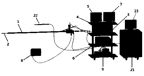
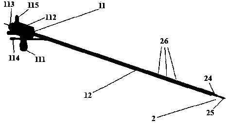
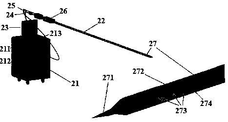
[0026] Ultrasound microscopy system for mediastinal lesions according to the present invention, such as figure 1 As shown, it includes: an ultrasonic rigid bronchoscope, which is composed of an improved rigid bronchoscope 1 , a catheter-type ultrasonic probe 2 , a cold light source host 3 , a camera host 4 and an ultrasound host 6 . Rigid bronchoscope 1 comprises endoscope main body 11 and elongated operating end 12 (see figure 2 ). The catheter type ultrasonic probe 2 is installed on the head of the operation end 12 . The endoscope main body 11 is connected to the light source 8 through a light source interface 111 . T...


PUM

PropertyMeasurementUnit
Outer diameteraaaaaaaaaa
Thicknessaaaaaaaaaa
The inside diameter ofaaaaaaaaaa

Abstract

The invention belongs to the field of medical apparatus and instruments, and particularly relates to an ultrasonic microscopic examination system for mediastinal lesions. The ultrasonic microscopic examination system comprises an ultrasonic rigid bronchoscope and a vacuum aspiration biopsy instrument. The ultrasonic rigid bronchoscope comprises an improved rigid bronchoscope, a catheter type ultrasonic probe, a cold light source main unit, a photographic main unit and an ultrasonic main unit; the rigid bronchoscope comprises an endoscope body and a slim operating end, and the catheter type ultrasonic probe is mounted at the head of the operating end; the endoscope body is connected with a light source by a light source interface; the cold light source main unit is connected with the photographic main unit; the catheter type ultrasonic probe is connected to the ultrasonic main unit; the vacuum aspiration biopsy instrument comprises a power control main unit of a vacuum aspiration pump system and a puncture biopsy needle. The ultrasonic microscopic examination system has the advantages that the ultrasonic microscopic examination system can be easily and conveniently operated when the mediastinal lesions are diagnosed and treated by doctors, the diagnosis accuracy can be improved, the false negative rate can be reduced, the occurrence rate of infective complications can be decreased, the operative time can be shortened, and accordingly economic burden on patients and suffering of the patients can be relieved.

Description

technical field [0001] The invention belongs to the field of medical devices, and in particular relates to an ultrasonic microscopy system for mediastinal lesions. Background technique [0002] Because of the particularity of the anatomical structure of the mediastinum, the diagnosis of mediastinal lesions is a difficult point in clinical work. At present, bronchial fiberoptic needle aspiration biopsy (TBNA) has been carried out abroad, but TBNA only conducts unguided "blind puncture" based on preoperative imaging information, and there are still some serious complications such as pneumothorax, bleeding, and mediastinal emphysema. Dangerous, and because the working end is soft, effective operation cannot be performed, which may easily lead to missed diagnosis and incomplete operation. [0003] Rigid bronchoscope, also known as "ventilated bronchoscope" because it can keep the airway unobstructed during operation, has been used clinically for more than 100 years. With the a...

Claims


Application Information

Patent Timeline
IPC IPC(8): A61B8/12A61B10/04
Inventor 谭筱江张丽张湘东张睿翔
Owner NANFANG HOSPITAL OF SOUTHERN MEDICAL UNIV
Learn More