A method for detecting the presence of nanoparticles in organs and tissues

A nanoparticle and tissue technology, applied in the direction of material analysis using measurement of secondary emissions, can solve the problems of high cost and cumbersome nanoparticle methods, and achieve the effect of reducing difficulty, easy operation and simple technology

Active Publication Date: 2017-01-04
HARBIN INST OF TECH
View PDF2 Cites 0 Cited by
  • Summary
  • Abstract
  • Description
  • Claims
  • Application Information

AI Technical Summary

Problems solved by technology

[0003] The present invention aims to solve the problems of cumbersome and high-cost existing methods for detecting nanoparticles in organs and tissues, and provides a method for detecting whether there are nanoparticles in organs and tissues

Method used


Image

  • A method for detecting the presence of nanoparticles in organs and tissues
  • A method for detecting the presence of nanoparticles in organs and tissues
  • A method for detecting the presence of nanoparticles in organs and tissues

Examples


specific Embodiment approach 1

[0016] Specific embodiment one: the method for detecting whether there are nanoparticles in the organ tissue in this embodiment is carried out according to the following steps:

[0017] 1. Wash the organ tissue with PBS buffer, take 0.3-0.5g into a centrifuge tube, add 500μL RIPA lysate, and break;

[0018] 2. Add 500 μL RIPA lysate to the centrifuge tube, shake and mix well to obtain tissue fluid, and filter the tissue fluid with a filter to obtain filtrate;

[0019] 3. Take the filtrate and place it in a new centrifuge tube, extract it with saturated phenol for 1-3 times, discard the phenol layer, and obtain liquid A;

[0020] 4. Centrifuge liquid A at 14,000 g for 30 minutes, discard the supernatant, and obtain a precipitate;

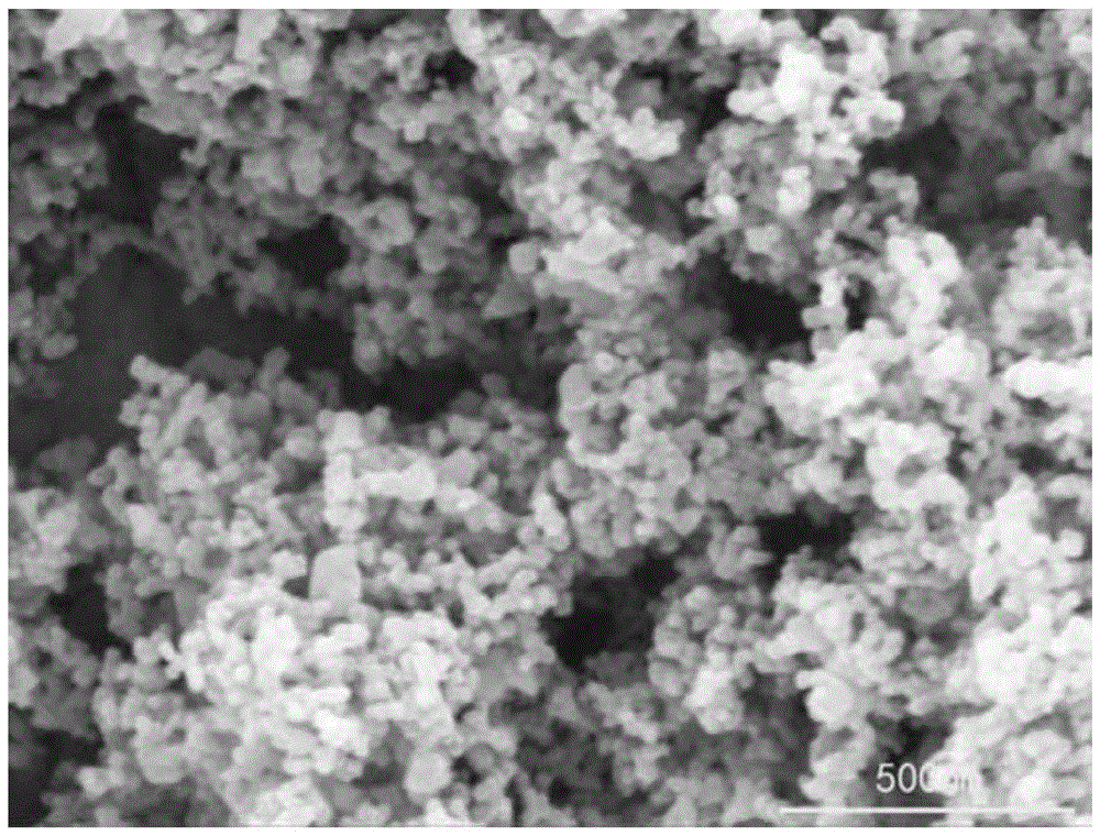
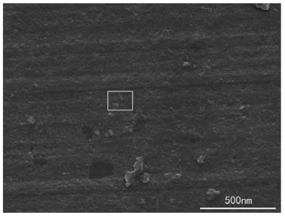
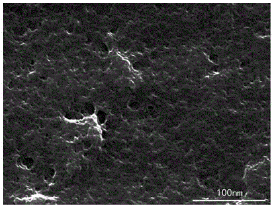
[0021] 5. Suspend the precipitate with 100-200 μL alcohol, drop it on the surface of the aluminum plate or copper plate, and perform scanning electron microscope detection after the alcohol evaporates;

[0022] 6. After the nanoparticles are observ...

specific Embodiment approach 2

[0024] Embodiment 2: The difference between this embodiment and Embodiment 1 is that the organs and tissues in Step 1 are fresh or frozen. Others are the same as in the first embodiment.

specific Embodiment approach 3

[0025] Embodiment 3: The difference between this embodiment and Embodiment 1 or 2 is that the crushing method in step 1 is ultrasonic crushing or mechanical stirring crushing. Others are the same as in the first or second embodiment.


PUM


Abstract

A method for detecting whether there are nanoparticles in organs and tissues relates to a method for detecting nanoparticles. The invention aims to solve the problems of cumbersome and high cost in the existing methods for detecting nanoparticles in organs and tissues. Method: 1. Wash the organ tissue, put it in a centrifuge tube, add the lysate to crush it; 2. Add the lysate, oscillate and mix to obtain the tissue fluid, filter the tissue fluid with a filter to obtain the filtrate; 3. Take the filtrate and put it in a new In a centrifuge tube, extract with saturated phenol, discard the phenol layer, and obtain liquid A; 4. Centrifuge liquid A to obtain a precipitate; Detection; 6. Carry out energy spectrum analysis to verify whether it is the nanoparticle to be detected. The method of the invention omits the processes of fixing, embedding and slicing, the sample preparation method is simpler and easier to operate, and does not require expensive slicing equipment. Used to detect the presence of nanoparticles in organs and tissues.

Description

technical field [0001] The invention relates to a method for detecting nanoparticles. Background technique [0002] With the development of nanotechnology, nanomaterials have been applied in various fields closely related to human beings, such as pharmaceuticals, food additives, papermaking, sewage treatment, etc. However, a large number of studies have shown that eating or contacting these nanomaterials for a long time will have negative Therefore, nano-toxicity has become a key topic today. At present, the main point of view on the mechanism of the direct harm of nanomaterials to the body is that when nanomaterials enter the organs in the form of particles, their nanoscale volume and specific surface area will cause a large accumulation of reactive oxygen species in the cells of the organs, resulting in reactive oxygen species. Oxygen metabolism disorder eventually leads to oxidative stress, and oxidative stress is an important threat to cells, which can cause DNA damage,...

Claims


Application Information

Patent Timeline
Patent Type & Authority Patents(China)
IPC IPC(8): G01N23/22
Inventor 顾宁马卓胡海龙郭倩
Owner HARBIN INST OF TECH
Why Eureka
Try Eureka