A method for judging coplanarity between two-dimensional ultrasound image and puncture needle

A technology of ultrasonic images and puncture needles, which is applied in ultrasonic/sonic/infrasonic diagnosis, sonic diagnosis, infrasonic diagnosis, etc. It can solve problems such as difficulty in judging the position of puncture needle, manual judgment of puncture needle, and affecting the accuracy of puncture needle.

Active Publication Date: 2016-06-29
HARBIN ENG UNIV
View PDF4 Cites 2 Cited by
  • Summary
  • Abstract
  • Description
  • Claims
  • Application Information

AI Technical Summary

Problems solved by technology

[0005] Second, it is necessary to manually judge the position of the puncture needle on the ultrasound image
Although the needle tip appears as a bright area on the ultrasound image, the brightness of the needle body is lower than that of the needle tip, and the bright spot in the surrounding tissue easily obliterates part of the needle body, making it difficult to determine the position of the puncture needle on the image. Therefore, the operator Subjective judgment will affect the correctness of the image coordinate positioning of the puncture needle

Method used


Image

  • A method for judging coplanarity between two-dimensional ultrasound image and puncture needle
  • A method for judging coplanarity between two-dimensional ultrasound image and puncture needle
  • A method for judging coplanarity between two-dimensional ultrasound image and puncture needle

Examples


Embodiment Construction

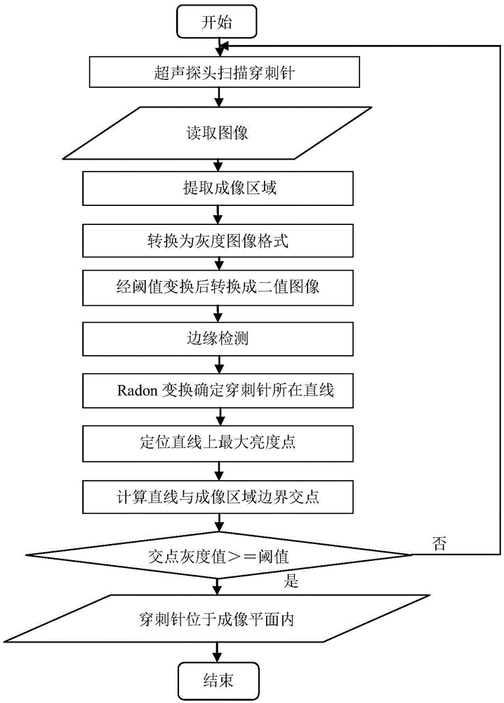
[0035] Such as figure 1 As shown, the method for judging the coplanarity between the two-dimensional ultrasonic image and the puncture needle of the present invention includes the following steps:

[0036] Step 1: Image preprocessing. Extract the imaging area on the ultrasound image, convert the color image into a grayscale image, and convert it into a binary image after threshold transformation;

[0037] Extract the imaging area on the ultrasound image: for a fan-shaped probe, extract a fan-shaped area, and for a rectangular probe, extract a rectangular area. The coordinates of these regions are determined by the parameter settings of the ultrasound imager.

[0038] Convert color images to grayscale images: Although the saved ultrasound images display grayscale objects, the image format is often stored as a color image, which is not convenient for further processing, so it is necessary to convert the image after extracting the imaging area from a color format in grayscale ...


PUM


Abstract

The present invention relates to a method for judging the coplanarity between a two-dimensional ultrasonic image and a puncture needle. A hand-held ultrasonic probe scans the puncture needle in the tissue, and saves the ultrasonic image of the puncture needle in the tissue. It is characterized in that: step 1: extracting the imaging area on the ultrasonic image, Convert the color image to a grayscale image, and convert it into a binary image after threshold transformation; Step 2: Perform edge detection on the binary image obtained in Step 1, and determine the straight line where the puncture needle is located through Radon transformation; Step 3: Locate the puncture needle The position of the needle tip; step 4: determine whether the puncture needle is located in the imaging plane by judging whether the gray value of the intersection point of the straight line where the puncture needle is located and the boundary of the ultrasound imaging area is greater than a certain threshold.

Description

technical field [0001] The invention relates to a method for judging the coplanarity between a two-dimensional ultrasonic image and a puncture needle. Background technique [0002] The image-guided surgery system uses medical imaging and computer graphics image technology to perform three-dimensional reconstruction and visualization of multi-modal image data of patients before surgery, obtain a three-dimensional model, formulate reasonable and quantitative surgical plans, and carry out preoperative simulation; Use the 3D space positioning equipment to register the image and the patient's physical space, map the patient's actual body position and the real-time spatial position of the surgical instrument to the patient's 3D image space, and collect and display the position of the surgical instrument in real time. Observe the relative positional relationship between the surgical instrument and the lesion in the three-dimensional image, and perform precise surgical treatment on ...

Claims


Application Information

Patent Timeline
Patent Type & Authority Patents(China)
IPC IPC(8): A61B8/00A61B90/00
Inventor 栾宽李金丛珊李海波孙小越
Owner HARBIN ENG UNIV
Why Eureka
Try Eureka