Coincidence SPECT tumor imaging quantitative analysis technology and application in tumor assessment

A CT image, tumor technology, applied in echo tomography, computed tomography scanner, etc., can solve problems such as qualitative analysis but not quantitative analysis.

Inactive Publication Date: 2015-05-13
刘丽 +1
View PDF0 Cites 10 Cited by
  • Summary
  • Abstract
  • Description
  • Claims
  • Application Information

AI Technical Summary

Problems solved by technology

[0004] The purpose of the present invention is to provide a method for Coincidence SPECT or SPECT/CT to quantitatively measure tumor SUV, SUL, MTV and TLG, to overcome th...

Method used


Image

  • Coincidence SPECT tumor imaging quantitative analysis technology and application in tumor assessment
  • Coincidence SPECT tumor imaging quantitative analysis technology and application in tumor assessment
  • Coincidence SPECT tumor imaging quantitative analysis technology and application in tumor assessment

Examples


Embodiment Construction

[0025] The invention provides an image quantitative analysis technology for tumor Coincidence SPECT imaging, which can obtain tumor tissue for 18 The uptake criteria of F-FDG imaging drugs, including SUV, SUL, MTV and TLG, which were previously only obtained in PET or PET / CT, can also be obtained in Coincidence SPECT or SPECT / CT through the new technology of the present invention accomplish.

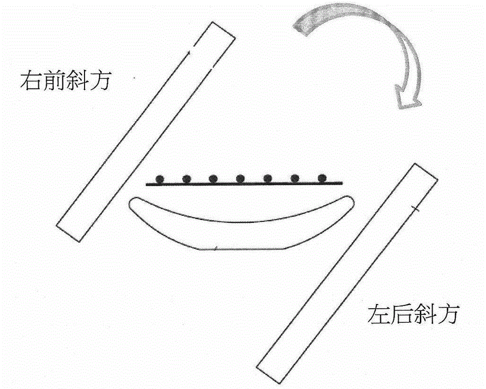
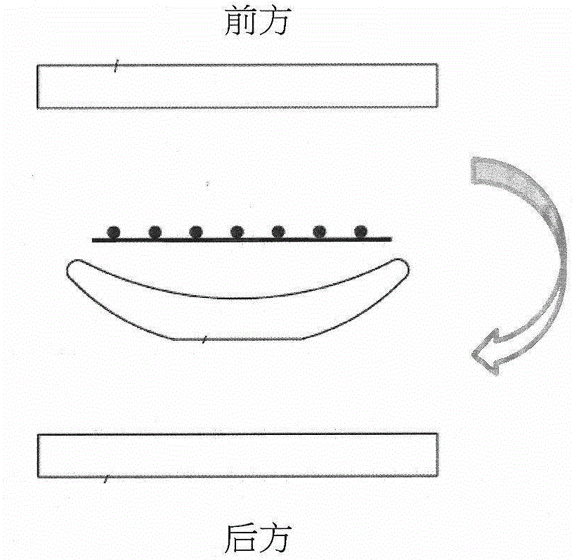
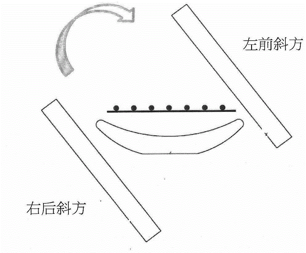
[0026] First, in the image acquisition step, use Coincidence SPECT or SPECT / CT to acquire images of the patient's tumor. Patients injected with F-18 labeled 18 After F-FDG tumor imaging drugs, start Coincidence SPECT or SPECT / CT dual probes and rotate 180 degrees to collect image data. In rotation, the original image data is represented by spatial coordinates and rotation angles. The probe can be rotated and collected from right anterior oblique to left posterior oblique, anterior to posterior, left anterior oblique to right posterior oblique, and posterior to anterior ( 1A to 1D ). ...


PUM


Abstract

The invention relates to a tumor imaging quantitative detection technology, in particular to a method for quantitatively measuring a tumor standard uptake value, a thin body standard uptake value, a metabolic volume and total glucolysis through Coincidence SPECT or SPECT/CT and application of the method in tumor assessment. The implementation steps of the method comprise an image acquisition step, a scatter correction step, a nuclide physical decay correction step, a movement correction step, a tissue attenuation correction step, an image spatial resolution recovery step, a noise removal step, a standard uptake value calculation step and a tumor therapeutic effect assessment step. By means of the technological means of the Coincidence SPECT tumor imaging quantitative analysis technology and the application in tumor assessment, the clinical problem of quantitatively measuring the tumor standard uptake value, the thin body standard uptake value, the metabolic volume and the total glucolysis is solved through the Coincidence SPECT and the SPECT/CT, and the technology can be used in the clinical assessment of tumor.

Description

【Technical field】 [0001] The invention relates to a quantitative detection technology of tumor images, in particular to a line-compliant single photon emission computed tomography (coincidence single photon emission computed tomography, Coincidence SPECT) or a line-compliant single photon emission computed tomography Quantitative measurement of tumor uptake by imaging / X-ray computed tomography (Coincidence SPECT / CT), including: standardized uptake value (SUV), standardized uptake lean body mass (SUL), metabolic volume (metabolic volume) volume, MTV) and total lesion glycolysis (TLG) measurement techniques, and the use of this technique in tumor assessment. 【Background technique】 [0002] SUV, SUL, MTV and TLG are currently clinically used fluorine-18 (F-18) labeled deoxyglucose (fludeoxyglucose, 18 F-FDG) imaging agent is the most important indicator for evaluating the uptake ability of imaging drugs by tumors, and has important clinical significance for tumor identificatio...

Claims


Application Information

Patent Timeline
IPC IPC(8): A61B6/03
Inventor 许百灵戴浩洁郑玉民
Owner 刘丽
Why Eureka
Try Eureka