Analytical processing method and device of medical image data

A medical image, analysis and processing technology, applied in the field of medical image data processing, to achieve comprehensive tracking, precise tracking, and accurate medical diagnosis results

Inactive Publication Date: 2015-07-01
SHANGHAI UNITED IMAGING HEALTHCARE
View PDF5 Cites 18 Cited by
  • Summary
  • Abstract
  • Description
  • Claims
  • Application Information

AI Technical Summary

Problems solved by technology

[0006] The problem solved by the present invention is that it is impossible to clearly, quickly, and concisely provide doctors with various interrelated medical image data at multiple time points, and it is impossible to accurately and conveniently analyze the medical image data, which in turn leads to the inability to accurately and timely diagnose patients and treatment issues

Method used


Image

  • Analytical processing method and device of medical image data
  • Analytical processing method and device of medical image data
  • Analytical processing method and device of medical image data

Examples


Embodiment 1

[0056] In this embodiment, an example of sorting two sets of adjacent medical image data sets among multiple sets of medical image data sets sorted by time is used for illustration.

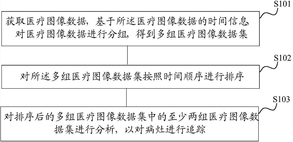
[0057] figure 2 It is a schematic flow chart of the analysis and processing method of medical image data provided in this embodiment, as figure 2 As shown, step S201 is firstly executed to obtain medical image data, and based on the time information of the medical image data, the medical image data is grouped to obtain multiple groups of medical image data sets. Please refer to step S101.

[0058] Step S202, sorting the multiple sets of medical image data sets in chronological order. Please refer to step S102.

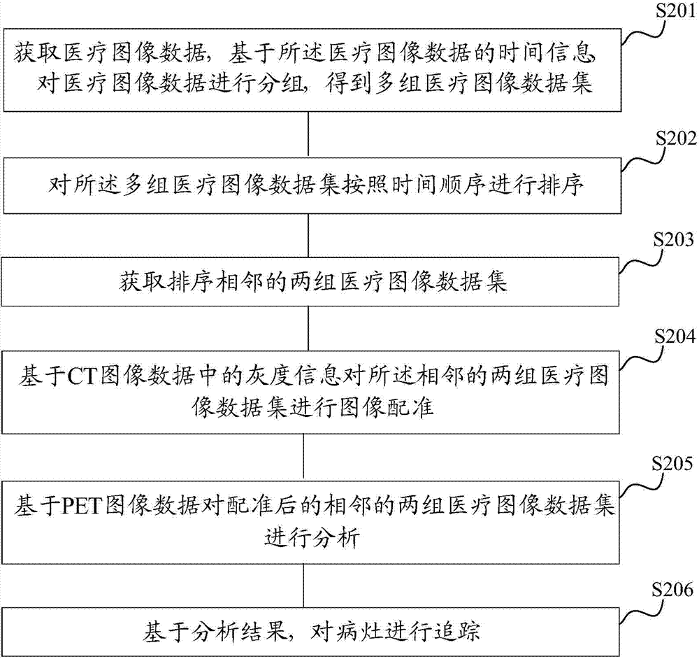
[0059] Step S203, acquiring two sets of medical image data sets that are sorted adjacent to each other.

[0060] Based on the multiple sets of chronologically ordered medical image datasets obtained in step S202, two sets of medical image datasets that are temporally adjacent are se...

Embodiment 2

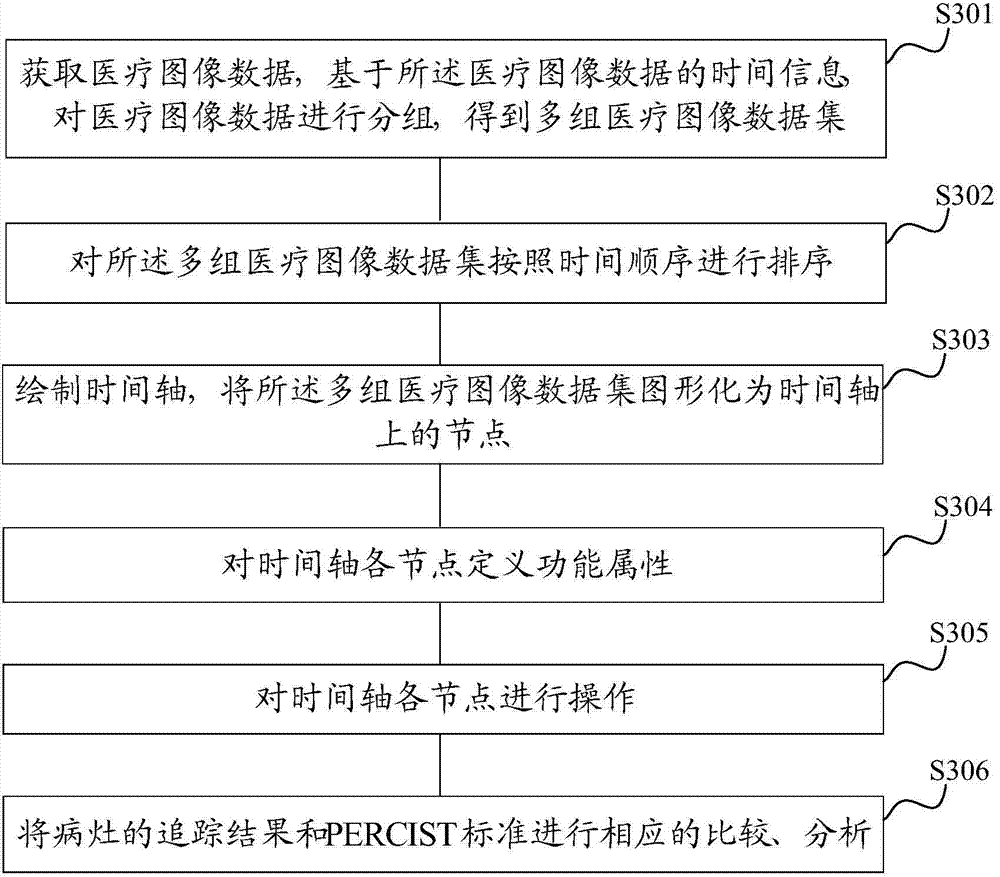
[0075] In this embodiment, a method for analyzing and processing medical image data based on the time axis is provided, which can represent medical image data scanned at different time points and in different medical imaging modes through different nodes on the time axis , and then by performing corresponding operations on different nodes on the time axis, the tracking of lesions is realized, and the tracking results of lesions are compared and analyzed with the PERCIST standard to realize accurate diagnosis of lesions.

[0076] image 3 It is a schematic flow chart of the analysis and processing method of medical image data provided in this embodiment, as image 3 As shown, step S301 is firstly executed to obtain medical image data, and based on the time information of the medical image data, the medical image data is grouped to obtain multiple groups of medical image data sets.

[0077] For step S301, please refer to step S101.

[0078] Step S302 is executed to sort the mu...

Embodiment 3

[0095] In this embodiment, the analysis and processing method of medical image data based on the time axis is adopted, as shown in the second embodiment Figure 4 As shown in the display interface, the timeline management template is as follows Figure 5 shown. In the process of image registration for adjacent nodes, a combination of automatic registration and manual registration is used for image registration. After the lesion is tracked, the tracking results of the lesion will be compared with the PERCIST standard. Comparison, analysis, and accurate diagnosis of the lesion.

[0096] Image 6 It is a schematic flow chart of the analysis and processing method of medical image data provided in this embodiment, as Image 6 As shown, step S601 is firstly executed to obtain medical image data, and based on the time information of the medical image data, the medical image data is grouped to obtain multiple groups of medical image data sets.

[0097] Step S602 is executed to sor...


PUM


Abstract

The invention discloses an analytical processing method and device of medical image data. The method comprises the following steps: obtaining medical image data, and grouping the medical image data on the basis of the time information of the medical image data to obtain multiple groups of medical image data sets, wherein the same group of medical image data set owns the same time information; sorting the multiple groups of medical image data sets according to a time sequence; and analyzing at least two groups of medical image data sets in multiple groups of sorted medical image data sets to trace focuses. The method can effectively process the medical image data at multiple time points, so that the complex medical image data can be mutually related, doctors can clearly, quickly and conveniently obtain the mutually-related medical image data so as to conveniently check, compare and analyze multiple groups of medical image data at different time points, and finally, the focuses can be accurately traced on the basis of an analysis result.

Description

technical field [0001] The invention relates to the field of medical image data processing, in particular to a method and device for analyzing and processing medical image data. Background technique [0002] With the continuous development of computer science and information technology, medical imaging technology has also been developed rapidly, and various medical imaging devices are constantly emerging. Such as computed tomography (CT, Computed Tomography), magnetic resonance imaging (MR, Magnetic Resonance), positron emission tomography (PET, Positron Emission Tomography), digital X-ray imaging (DR, Digital Radiography) and single photon emission computed tomography Imaging (SPECT, Single-Photon Emission Computed Tomography), etc. [0003] However, various imaging techniques and inspection methods have their advantages and disadvantages. Not one imaging technique can be applied to the examination of all organs of the human body and the diagnosis of diseases, nor can one ...

Claims


Application Information

Patent Timeline
Patent Type & Authority Applications(China)
IPC IPC(8): G06F19/00G06T7/00
Inventor 程兆宁王季勇钱政
Owner SHANGHAI UNITED IMAGING HEALTHCARE
Why Eureka
Try Eureka