Pelvic organ automatic segmentation method for CT examination

An automatic segmentation and organ technology, applied in the field of image processing, can solve the problems of insufficient soft tissue contrast, time-consuming work of organ contour outline, and inability to apply clinical treatment, etc.

Active Publication Date: 2015-12-23
SHANDONG NORMAL UNIV
View PDF2 Cites 6 Cited by
  • Summary
  • Abstract
  • Description
  • Claims
  • Application Information

AI Technical Summary

Problems solved by technology

This has led to time-consuming work for doctors to delineate the organ contours in prostate CT, and the instability of manual delineation will lead to some unpredictable problems in the process of accurate treatment
In recent years, although there have been some methods for segmenting prostate CT to outline organs, these methods are currently unavailable due to the lack of soft tissue contrast in CT images and the variability of pelvic organ morphology mentioned above. to clinical treatment

Method used


Image

  • Pelvic organ automatic segmentation method for CT examination
  • Pelvic organ automatic segmentation method for CT examination
  • Pelvic organ automatic segmentation method for CT examination

Examples


Embodiment Construction

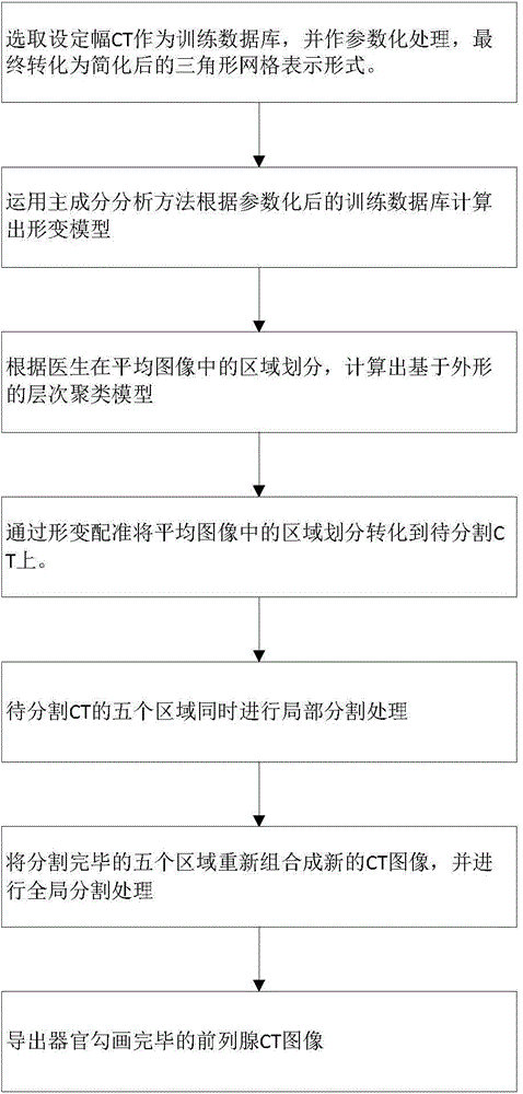
[0091] The present invention will be further described below in conjunction with the accompanying drawings and embodiments.

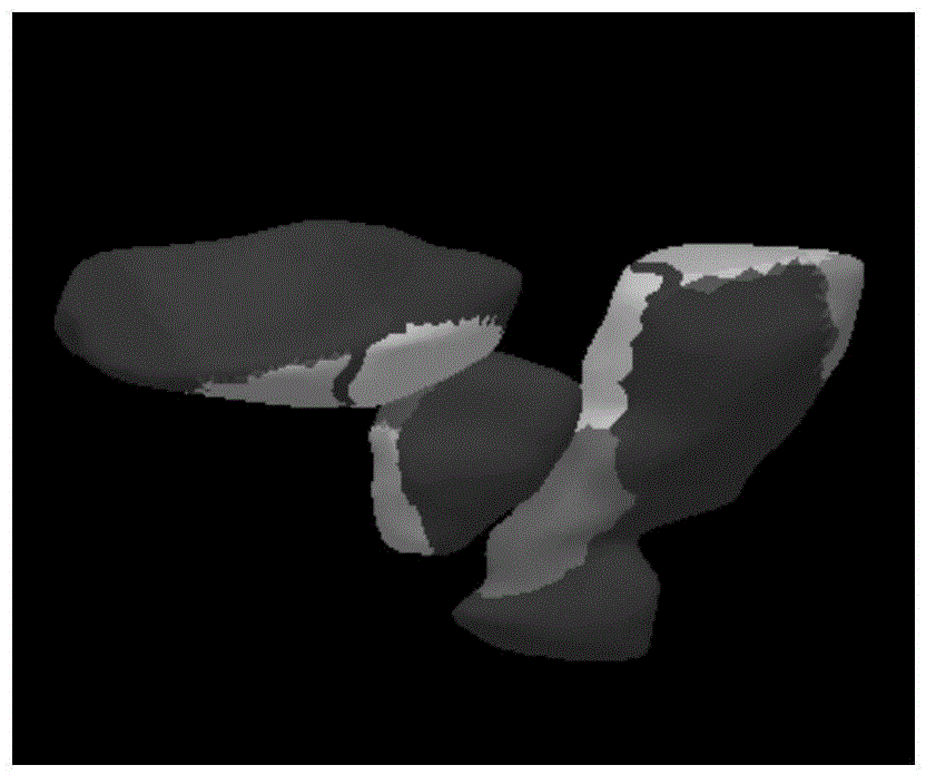
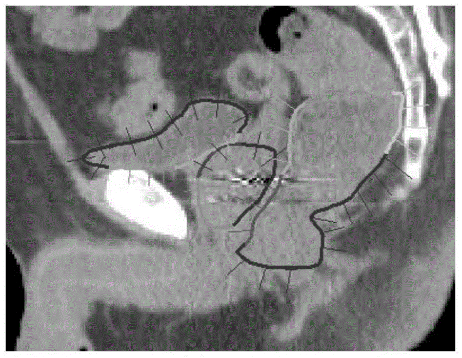
[0092] Using a population-based prostate CT database, a deformation model to guide segmentation and a hierarchical clustering model to enhance segmentation accuracy were established. Among them, the establishment of the deformation model uses the method of principal component analysis and multivariate statistics; while the establishment of the hierarchical clustering model based on the shape is divided into five regions according to the anatomical structure and deformation characteristics of the pelvic organs, and each region is established separately. Hierarchical clustering models for ease of use. In the segmentation process, the deformable model is used as the contour guidance, and the best segmentation contour of the region is found according to the hierarchical clustering model of each region, and then the contours of the five regions are recombine...


PUM


Abstract

The invention discloses a pelvic organ automatic segmentation method for CT examination. The method comprises the steps of based on the model establishment process of a training database, establishing a deformable model for the segmentation process and hierarchical clustering models, wherein the hierarchical clustering models are formed in such a manner that organs in the training database are segmented into five regions for the amplitude-stated prostate CT examination and each region is in the form of an outline-based hierarchical clustering model established according to the segmentation of the amplitude-stated prostate CT examination; during the local segmentation process, adopting the deformable model as a deformation guidance, finding out an optimal segmentation outline for each region according to the hierarchical clustering model of the region, and recombining the optimal segmentation outlines of the five regions as an initial outline of the global segmentation process; during the global segmentation process, segmenting and regulating the initial outline of the global segmentation process in the same way with that of the local segmentation process to obtain final segmented images.

Description

technical field [0001] The invention relates to the field of image processing, in particular to a method for automatic segmentation of pelvic organs in CT. Background technique [0002] In today's society, due to unhealthy living habits, prostate disease has become one of the main diseases that plague men. In the treatment of prostate diseases, 3D-CT of the prostate is one of the main means for doctors to obtain the specific conditions of patients, and it is also the main reliance to improve the treatment effect and treatment accuracy in the process of clinical treatment. In order to use prostate CT for accurate clinical treatment, the contours of each tissue must be accurately delineated, and this process is generally completed manually by doctors. [0003] However, this process takes about 15-20 minutes, and the outlines of tissues and organs in the same CT image vary greatly between different doctors. Prostate CT is mainly divided into three parts: prostate, bladder, an...

Claims


Application Information

Patent Timeline
Patent Type & Authority Applications(China)
IPC IPC(8): G06T7/00
CPCG06T7/0012G06T2207/10081G06T2207/30081
Inventor 李登旺臧鹏霄柴象飞张路路李洪升尹勇陈进琥马长升
Owner SHANDONG NORMAL UNIV
Why Eureka
Try Eureka