Medical application of creg protein in protecting myocardial ischemia-reperfusion injury
A technology for reperfusion injury and myocardial ischemia, which can be used in medical preparations containing active ingredients, pharmaceutical formulas, peptide/protein components, etc., and can solve problems such as unclearness
- Summary
- Abstract
- Description
- Claims
- Application Information
AI Technical Summary
Problems solved by technology
Method used
Image
Examples
Embodiment 1
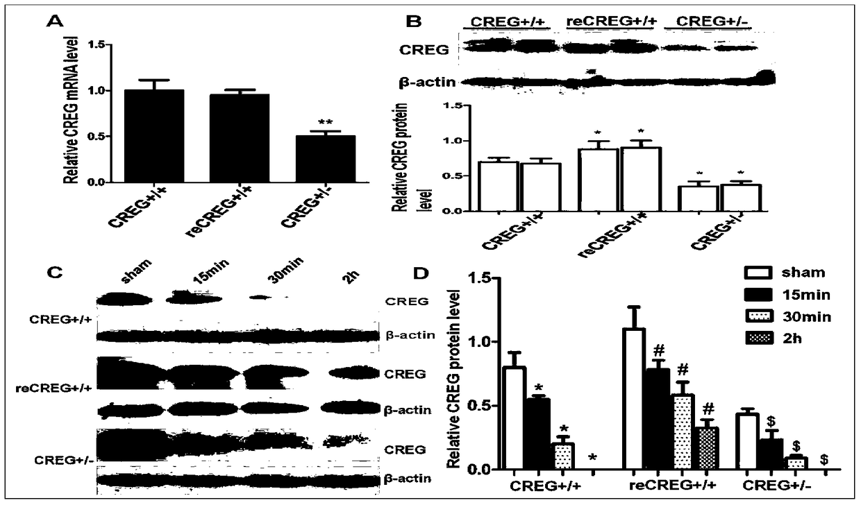
[0052] Example 1. Giving mice myocardial ischemia-reperfusion treatment can down-regulate the expression of CREG protein in the myocardium
[0053] ①Establishment of myocardial ischemia-reperfusion model in mice
[0054] Prepare various instruments, materials and medicines before the operation; the steps and methods of thoracotomy and ligation of the left anterior descending coronary artery: cut the skin 2 mm to the left side of the sternum, bluntly separate the muscles to see the ribs, and gently use ophthalmic scissors in the fourth intercostal space The intercostal muscles were separated downward, and the vascular forceps were extended upward to repeatedly clamp the two ribs to reduce bleeding. Cut ribs 3 and 4, pull apart the chest wall with retractors, thread a pair of threads on the chest wall muscles on both sides and leave small loops on the inside. Carefully cut the pericardium and apply gentle pressure to the heart with a cotton swab. Insert the needle at about 2 m...
Embodiment 2
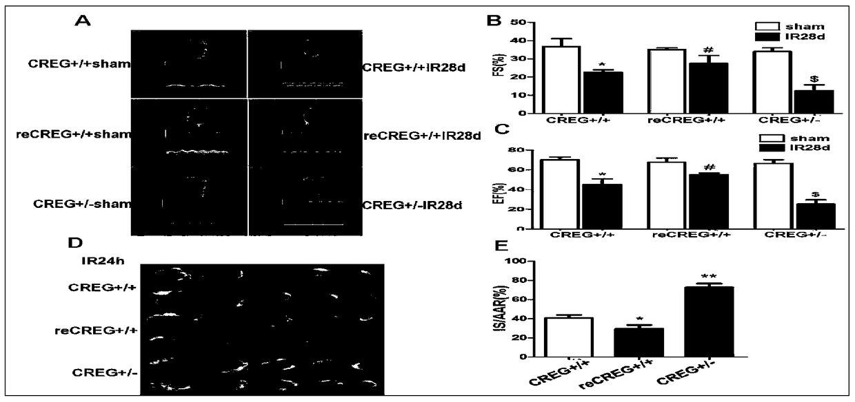
[0073] Example 2. Changes in the expression of CREG protein in mouse myocardium can affect the heart function of mice
[0074] ① CREG at 28 days after myocardial ischemia-reperfusion + / + and CREG + / - Comparison of heart function. The Vevo2100 ultrasonic biomicroscope imaging system from VisualSonics Corporation of Canada was adopted, and the center frequency of the probe was 30MHz. After the mice were anesthetized with 2% isoflurane, the hair on the chest and abdomen was removed. The mice were fixed in a supine position on a constant temperature inspection table, and the temperature was kept at 37°C. The physiological parameters such as ECG and respiration were recorded synchronously. The heart rate was maintained at about 450 beats / min. After the heart rate was stable for 1 min, smear coupling agent on the chest and perform ultrasound biomicroscopy. examine. The results showed that after 28 days of myocardial ischemia-reperfusion, CREG + / - CREG + / + The cardiac function ...
Embodiment 3
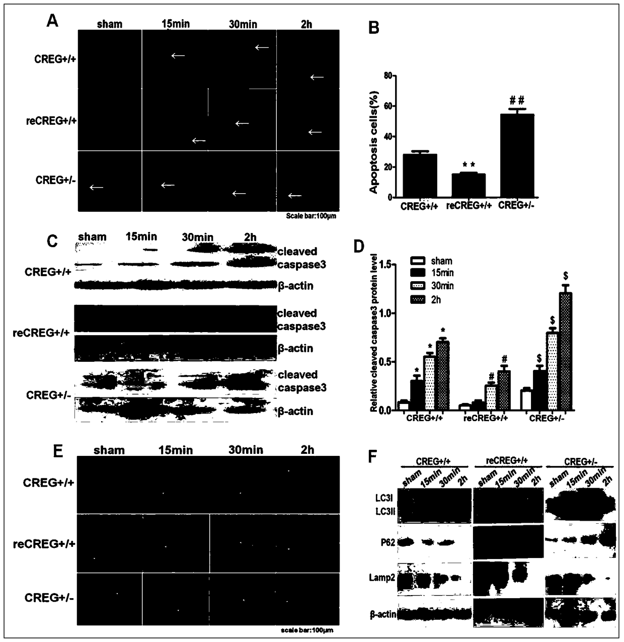
[0078] Example 3. Administration of exogenous recombinant CREG protein during myocardial ischemia-reperfusion can reduce the apoptosis of mouse cardiomyocytes and further activate the occurrence of autophagy
[0079] ①The apoptosis of cardiomyocytes in the myocardium of three kinds of mice was detected by Tunel staining. After preparing the necessary reagents for Tunel staining, the frozen tissue sections were fixed in 4% paraformaldehyde at room temperature for 10 min, and washed twice with PBS, 10 min each time. After 20 minutes with 3% hydrogen peroxide methanol solution, wash with PBS 3 times, 5 minutes each time, place on ice with 0.1% sodium citrate for 2 minutes, add Tunel staining solution to the surface of the sample, and incubate in a 37°C incubator for 1 hour, protected from light; soak in PBS , DAPI stained nuclei. The results suggest that after myocardial ischemia-reperfusion CREG + / - The mice had the most cardiomyocyte apoptosis, while the mice given exogenous ...
PUM
 Abstract
Description
Claims
Application Information
 - R&D
- Intellectual Property
- Life Sciences
- Materials
- Tech Scout
- Unparalleled Data Quality
- Higher Quality Content
- 60% Fewer Hallucinations
Browse by: Latest US Patents, China's latest patents, Technical Efficacy Thesaurus, Application Domain, Technology Topic, Popular Technical Reports.
© 2025 PatSnap. All rights reserved.Legal|Privacy policy|Modern Slavery Act Transparency Statement|Sitemap|About US| Contact US: help@patsnap.com


