Fusion protein molecular weight analysis method

A technology of fusion protein and analysis method, applied in the direction of biological testing, material inspection products, etc.

Active Publication Date: 2016-06-01
SUNSHINE GUOJIAN PHARMA (SHANGHAI) CO LTD
View PDF1 Cites 2 Cited by
  • Summary
  • Abstract
  • Description
  • Claims
  • Application Information

AI Technical Summary

Problems solved by technology

[0003] Glycosylation modification is very important to the biological function of fusion protein, but complex glycosy

Method used


Image

  • Fusion protein molecular weight analysis method
  • Fusion protein molecular weight analysis method
  • Fusion protein molecular weight analysis method

Examples


Embodiment 1

[0033] Example 1: Analysis of the complete molecular weight of the fusion protein (removal of N-sugar first, and then removal of sialic acid)

[0034] Fusion protein complete molecular weight analysis steps:

[0035] (1) Removal of N-Glycosylations

[0036] Get 50 μ g Etanercept fusion protein sample (Pfizer Pharmaceutical Co., Ltd., the same below), add 50 mM NH 4 HCO 3 (pH8.0) solution to a volume of 20 μL, add 1 μL of 4-fold diluted peptide N-glycosidase PNGaseF (NEB, 500000u / mL), mix well, and incubate at 37°C for 24 hours.

[0037] (2) Sialic acid removal

[0038] For the sample incubated in (1), adjust the pH value to 5-6 with 1% FA, add 0.5 μL of sialidase Neuraminidase (Roche, 1 U / 100 μL), and incubate at 37° C. for 24 hours.

[0039] (3) ESI-Q-TOFMS analysis

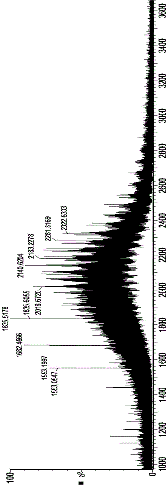
[0040] The fusion protein of asialo de-N-glycans adopts 50mM NH 4 HCO 3 (pH8.0) solution was diluted to 1 mg / mL; then MassPREP™ MicroDesaltingColumn 2.1 × 5mm desalting column (Waters, the same below) aft...

Embodiment 2

[0042] Example 2: Analysis of the complete molecular weight of the fusion protein (sialic acid is removed first, and then N-sugar is removed)

[0043] Fusion protein complete molecular weight analysis steps:

[0044] (1) Sialic acid removal

[0045] Take 50μg Etanercept fusion protein sample, add 50mM NH 4 HCO 3 (pH 8.0) solution to a volume of 20 μL, adjusted the pH value to 5-6 with 1% FA, added 0.5 μL of sialidase Neuraminidase, and incubated at 37° C. for 24 hours.

[0046] (2) N-sugar removal

[0047] The sample incubated in (1), with 1% NH 3 .H 2 O to adjust the pH value to 7-8, add 1 μL of 4-fold diluted PNGaseF (NEB, 500000u / mL), and incubate at 37°C for 24 hours.

[0048] (3) ESI-Q-TOFMS analysis

[0049] De-N-sugar asialo fusion protein using 50mM NH 4 HCO 3 (pH8.0) solution was diluted to 1mg / mL; then MassPREPTMMicroDesaltingColumn2.1 × 5mm desalting column desalting, and finally mass spectrometry analysis.

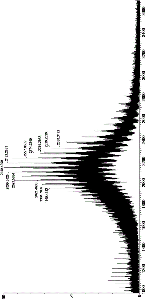
[0050] Experimental results: See the attachment...

Embodiment 3

[0051] Example 3: Comparison of molecular weight analysis of asialo deconvoluted intact protein under different pH conditions after N-sugar removal

[0052] Fusion protein complete molecular weight analysis steps:

[0053] (1) N-sugar removal

[0054] Take 400μg Etanercept fusion protein sample, add 50mM NH 4 HCO 3 (pH8.0) solution to a volume of 160 μL, add 2 μL PNGaseF, mix well, and incubate at 37°C for 24 hours.

[0055] (2) Sialic acid removal

[0056] Take 4 samples incubated in (1), 50 μg each, adjust the pH to 4, 5, 6, and 7 with 1% FA, add 0.5 μL of sialidase Neuraminidase, and incubate at 37°C for 24 hours.

[0057] (3) ESI-Q-TOFMS analysis

[0058] The fusion protein of de-N-sugar and asialic acid uses 50mM NH 4 HCO 3 (pH8.0) solution diluted to 1mg / mL; then MassPREPTMMicroDesaltingColumn2.1 × 5mm after desalting, mass spectrometry.

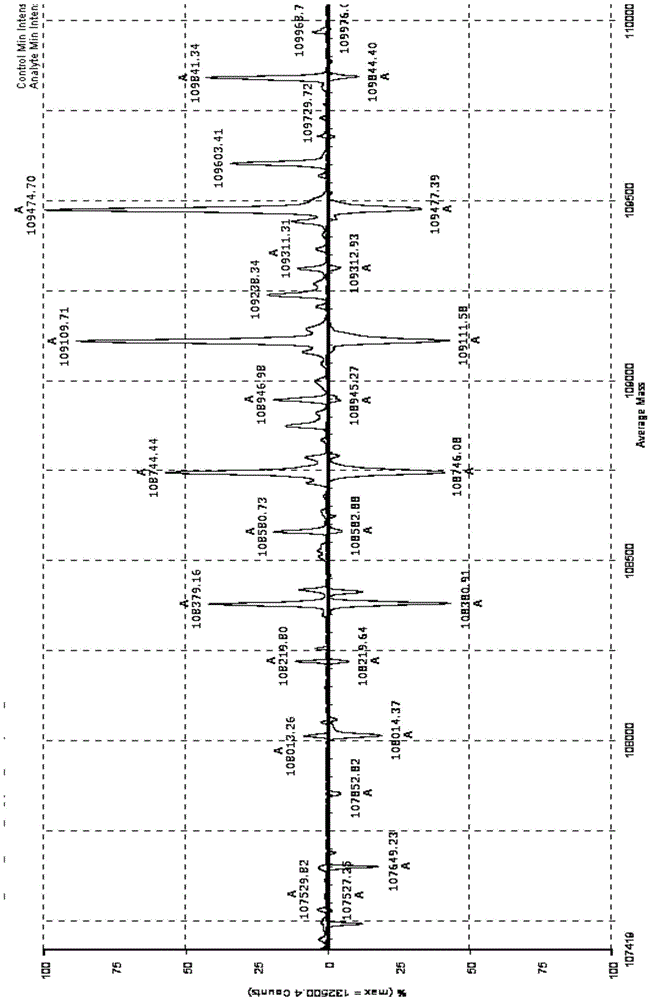
[0059] Experimental results: After N-sugar removal, the molecular weight comparison analysis results of asialic acid deconvol...


PUM


Abstract

The present invention discloses a fusion protein molecular weight analysis method, and specifically relates to a method for carrying out complex glycosylated fusion protein accurate molecular weight analysis by using ESI-Q-TOF, particularly to a sample pre-treatment method for removing fusion protein N-glycan and sialic acid by using specific glycan removing enzyme, wherein the treated fusion protein is separated through a salt removing column, the effluent is broken through an ESI source to obtain continuous multi-charged ions, and tandem Q-TOF mass spectrometry analysis and deconvolution calculation are performed to obtain the complete protein accurate molecular weight of the O-glycan core isomer. With the method of the present invention, the high inhibition of the N-glycan and/or sialic acid on the fusion protein ionization efficiency can be overcome, and the complete protein molecular weight of the O-glycan fusion protein can be accurately determined.

Description

technical field [0001] The invention belongs to the technical field of biomedicine, and in particular relates to a protein molecular weight analysis method. Background technique [0002] Cytokine recombinant fusion protein is a kind of gene sequence that uses genetic engineering to connect coding cytokines and other protein molecules with specific functions and express the corresponding protein fusion products. Its structural feature is that the functional domain of cytokines is fused with the active domains of other molecules, and each component can play a synergistic effect, so that the biological activity of the fusion protein is greatly enhanced compared with the monomers. Glycosylation modification of fusion protein is closely related to its biological activity, structure and pharmacokinetics, and will also directly or indirectly affect its resistance to proteases, binding to Fc receptors, interaction with complement C1q components, and in vivo Half-life, relevant biol...

Claims


Application Information

Patent Timeline
IPC IPC(8): G01N33/68
Inventor 吕锋华刘周阳环民霞黄黎明谭青乔
Owner SUNSHINE GUOJIAN PHARMA (SHANGHAI) CO LTD
Why Eureka
Try Eureka