Laparoscopic uterine fibroid crushing bag

A uterine fibroids and laparoscopy technology, applied in the field of medical devices, can solve the problems of malignant tumor cell dissemination, distant spread of malignant tumors, residual uterine fibroids cells, etc. Simple to use effects

Inactive Publication Date: 2016-06-29
李武
View PDF10 Cites 6 Cited by
  • Summary
  • Abstract
  • Description
  • Claims
  • Application Information

AI Technical Summary

Problems solved by technology

[0004] But the main defect that said method exists is: (1) in the course of operation, the electric cutter head of laparoscopic electric rotary cutter rotates and cuts, and unprotected uterine fragment tissue can be " launched " to each direction in abdominal cavity, makes smooth muscle Tumor cells plant and grow on the surface of the abdominal cavity, resulting in iatrogenic peritoneal uterine leiomy

Method used


Image

  • Laparoscopic uterine fibroid crushing bag

Examples


Embodiment Construction

[0024] The present invention will be described in detail below in conjunction with specific embodiments and accompanying drawings.

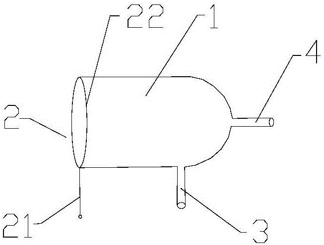
[0025] figure 1 It is a structural schematic diagram of a laparoscopic uterine fibroid crushing bag in an embodiment of the present invention. like figure 1 As shown, a laparoscopic uterine fibroid crushing bag, the crushing bag is divided into two parts, the main bag body 1 and the operating rod 2.

[0026] Wherein the main bag body 1 is a bag body with an open end. The main bag body in the present invention can be made of a variety of materials, such as medical plastics, medical silica gel, considering the opening size of the puncture hole, preferably thinner, and a plastic bag made of a material with higher strength, which can reduce the volume. It can also prevent damage.

[0027] In one embodiment of the present invention, the main bag body 1 is long cylindrical, such as figure 1 As shown, left end open. The cross section of the openin...


PUM

PropertyMeasurementUnit
Diameteraaaaaaaaaa
Diameteraaaaaaaaaa

Abstract

The invention provides a laparoscopic uterine fibroid crushing bag. The crushing bag is divided into two parts, namely a main bag body and an operating rod, wherein the main bag body is a bag body which is open in one end; the open end of the bag body is provided with two hollow arms, namely a first operating arm and a second operating arm on the bag body; the operating rod comprises an operating rod main body and a metal wire ring connected onto the operating rod main body; the metal wire ring is prepared from memory metal; and the metal wire ring is fixedly connected to the open rod the main bag body to form an inlet of the crushing bag. The laparoscopic uterine fibroid crushing bag is simple to operate, avoids the defect of the conventional laparoscopic uterine fibroid crushing, and completely keeps the minimally-invasive advantage of the laparoscopic hysteromyomectomy.

Description

technical field [0001] The invention belongs to the field of medical equipment, and relates to a pulverizing bag used for laparoscopic electric rotary cutting to pulverize hysteromyoma. Background technique [0002] Laparoscopic surgery is to insert a laparoscopic lens into the abdominal cavity, and use digital camera technology to transmit the images captured by the laparoscopic lens to the post-stage signal processing system through optical fibers, and display them on a dedicated monitor in real time. Then the doctor analyzes and judges the patient's condition through the images of different angles of the patient's organs displayed on the monitor screen, and uses special laparoscopic instruments to perform the operation. [0003] During laparoscopic myomectomy, when the diameter of myoma is larger than 20mm, the specimen must be pulverized with an electric rotary cutter. The electric rotary cutter can quickly and efficiently cut the fibroids into strips, compress and take...

Claims


Application Information

Patent Timeline
IPC IPC(8): A61B17/00A61B17/42
CPCA61B17/00234A61B17/4241A61B2017/00238A61B2017/00353A61B2017/4216
Inventor 李武
Owner 李武
Why Eureka
Try Eureka