Device and method for repeated immunostaining of same tissue section

A technology of tissue sectioning and multiple staining, which is applied in the field of medical biological experiment equipment, can solve the problems that immunostaining methods are difficult to implement, and achieve the effect that is conducive to analysis and diagnosis

Inactive Publication Date: 2016-07-20
NANCHANG DEMANDUO TECH CO LTD
View PDF3 Cites 8 Cited by
  • Summary
  • Abstract
  • Description
  • Claims
  • Application Information

AI Technical Summary

Problems solved by technology

If the study of the expression of multiple antigens is performed on a very small amount of tissue specimens (such as fine needle aspiration specimens), conventional immunostaining methods are difficult to achieve due to the limitation of specimen volume

Method used


Image

  • Device and method for repeated immunostaining of same tissue section
  • Device and method for repeated immunostaining of same tissue section

Examples


Embodiment Construction

[0012] The present invention will be described in detail below in conjunction with the accompanying drawings and specific examples.

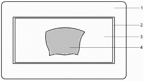
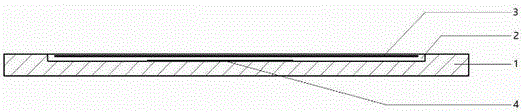
[0013] like figure 1 and figure 2 As shown, the tissue section carrying device includes a grooved glass slide 1 and a cover glass 3. The grooved glass slide 1 has a length of 60mm, a width of 40mm, and a thickness of 2.5mm except for the groove 2. , the bottom thickness of the groove 2 is 1.0mm; the length of the groove 2 is 42mm, the width is 26mm, and the depth is 1.5mm, and the inner wall of the groove is coated with polylysine; the length of the cover glass 3 is 40mm, and the width is 1.5mm. It is 24mm and the thickness is 0.13mm.

[0014] Using the tissue section carrying device to realize multiple immunostaining, the following two methods can be used:

[0015] Method 1: Routine paraffin-embedded tissue sections (thickness 4 μm) from lymph node puncture specimens were floated, and a tissue section 4 was picked up with a grooved glass sl...


PUM

PropertyMeasurementUnit
thicknessaaaaaaaaaa
lengthaaaaaaaaaa
widthaaaaaaaaaa

Abstract

The invention discloses a device and method for repeated immunostaining of the same tissue section in order to realize repeated immunostaining of the same tissue section and save tissue samples and meet diagnosis needs, including a tissue section bearing device and an application method thereof. The tissue section bearing device comprises a glass slide with a groove and a cover glass. The tissue section bearing device is adopted for repeated staining of sequentially circulating staining, photographing, cleaning, staining again, photographing again and cleaning again of the same tissue section; and through comparative analysis of pictures taken after the repeated staining, the antigen expression profile of each cell on the tissue section can be obtained.

Description

technical field [0001] The invention relates to a medical biological experiment device and method, in particular to a device and method for performing multiple times of immunostaining on the same tissue section. Background technique [0002] Immunostaining is to use the principle of specific combination of antigen and antibody to detect the expression of specific protein (antigen). It is one of the most commonly used detection methods to discover the functional changes of the body under physiological or pathological conditions. The most commonly used method is immunohistochemical staining. and immunofluorescence staining. The conventional method is to stain a tissue section with one antigen marker, but to study the expression of multiple antigens requires multiple tissue sections. If a very small amount of tissue specimens (such as fine needle aspiration specimens) is used to study the expression of multiple antigens, conventional immunostaining methods are difficult to ach...

Claims


Application Information

Patent Timeline
Patent Type & Authority Applications(China)
IPC IPC(8): G01N33/533
CPCG01N33/533
Inventor 不公告发明人
Owner NANCHANG DEMANDUO TECH CO LTD
Why Eureka
Try Eureka