High-precision three-dimensional chemical shift imaging method

A technology of chemical shift and imaging method, which is applied in the field of medical magnetic resonance imaging technology and imaging diagnostics, and can solve problems such as limited image resolution, phase sign calculation error, and unbalanced frequency encoding gradient.

Active Publication Date: 2016-07-20
谱影医疗科技(苏州)有限公司
View PDF5 Cites 18 Cited by
  • Summary
  • Abstract
  • Description
  • Claims
  • Application Information

AI Technical Summary

Problems solved by technology

However, in the case of unsatisfactory performance of the magnet and gradient system, not only the phase of the echo signal often exceeds the -π to +π range, causing serious phase winding artifacts, but also the phase of the water-lipid in the tissue area where the water-lipid coexists Signs are prone to miscalculations, resulting in incomplete water-fat separation; in particular, the use of a single-shot acquisition method based on bipolar (i.e., positive and negative) gradients for frequency encoding will Significantly increases the phase error
These problems have become the technical bottleneck for the routine application of the single-shot method in the clinical environment, reducing the diagnostic value of water-lipid separation images
In addition, the two-point and three-point Dxion imaging methods restrict the further improvement of image signal-to-noise ratio or scanning efficiency, and the two-dimensional scanning method used in conventional imaging also limits the image resolution in the layer selection direction

Method used


Image

  • High-precision three-dimensional chemical shift imaging method
  • High-precision three-dimensional chemical shift imaging method
  • High-precision three-dimensional chemical shift imaging method

Examples


Embodiment 1

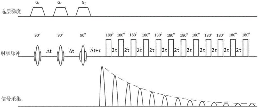
[0091] On the 0.35T medical magnetic resonance imager according to Figure 6Edit the imaging sequence as shown, set the area of ​​the first frequency encoding gradient to twice the area of ​​the pre-reading gradient, set the time interval between the center position of the RF excitation soft pulse and the center position of the first thick block selection gradient (that is, the first The echo time TE) is the minimum value, such as TE=6ms, set Δt slightly larger than the width of the frequency encoding gradient, such as Δt=2ms, set the polarity of the second frequency encoding gradient and the polarity of the first frequency encoding gradient Same, the two areas are equal, set Δτ=1 / Δf / 2=9.8ms according to the resonance frequency difference between water and fat protons Δf=51Hz, and set the layer selection gradient and phase encoding gradient according to the conventional spin echo sequence, and then save sequence file. In the sequence parameter table, set the size of the acqui...

Embodiment 2

[0117] Edited on a 1.5T medical magnetic resonance imager Figure 5 In the imaging sequence shown, the area of ​​the first frequency encoding gradient is set to be twice the area of ​​the pre-reading gradient, and the time interval between the center position of the radio frequency excitation soft pulse and the center position of the first thick block selection gradient is set (that is, the first cycle Wave time TE) is the minimum value, such as TE=3ms, set Δt slightly larger than the width of the frequency encoding gradient, such as Δt=1.6ms, set the polarity of the second frequency encoding gradient and the polarity of the first frequency encoding gradient Same, the two areas are equal, set the third and fourth frequency encoding gradient parameters in the same way, set Δτ=1 / Δf / 2=2.2ms in the sequence according to the chemical shift difference Δf of water and fat protons, and Set the layer selection gradient and phase encoding gradient according to the conventional spin echo...

Embodiment 3

[0148] Edited on a 1.5T medical magnetic resonance imager Figure 8 (or Figure 9 ), set the area of ​​the first frequency encoding gradient to be twice the area of ​​the pre-reading gradient integral, and set the time interval between the center position of the radio frequency excitation soft pulse and the center position of the first thick block selection gradient (that is, the first The first echo time TE) is the minimum value, such as TE=4ms, set Δt slightly larger than the width of the frequency encoding gradient, such as Δt=2ms, set the polarity of the second frequency encoding gradient and the polarity of the first frequency encoding gradient The properties are the same, the two areas are equal, set the third and fourth frequency encoding gradient parameters in the same way, set Δτ=1 / Δf / 2=2.2ms in the sequence according to the chemical shift difference Δf of water and fat protons, And refer to the conventional spin echo sequence to set the layer selection gradient and ...


PUM


Abstract

Disclosed in the invention is a high-precision three-dimensional chemical shift imaging method. A water image and a fat image that are separated from each other and in-phase graphs and anti-phase graphs of the water image and the fat image can be obtained by scanning once by using a three-dimensional single excited long echo string collection way, a radio frequency refocusing pulse string or frequency coding gradient technology, a pre scanning scheme having an echo amplitude and phase error precision correction feature, and a data post processing method. The method has the low requirements on magnetic field uniformity and gradient system performances; and the image quality and the diagnosis value can be improved obviously.

Description

technical field [0001] The invention relates to the technical fields of medical magnetic resonance imaging technology and image diagnostics, in particular to a high-precision three-dimensional chemical shift imaging method. Background technique [0002] In medical magnetic resonance imaging (MRI), fat-suppressed imaging is significantly superior to conventional imaging techniques in showing lesions, for example, it is beneficial to improve the sensitivity and specificity of diagnosing articular cartilage damage, and can improve the contrast of breast and optic nerve images. In T1-weighted images, fat is easily confused with contrast agent-enhanced lesions in enhanced scan due to short T1 and appears high signal, which affects the clinical diagnostic significance of enhanced scan. In addition, at 1.5T, the chemical shift frequency difference of water and fat is 210 Hz, and the RF bandwidth is mostly in the range of 1-3 kHz. Under the condition of 200 Hz / pixel, the water-fat ...

Claims


Application Information

Patent Timeline
Patent Type & Authority Applications(China)
IPC IPC(8): G01R33/56
CPCG01R33/56
Inventor 罗会俊
Owner 谱影医疗科技(苏州)有限公司
Why Eureka
Try Eureka