An ultrafast single-shot four-point dixon imaging method for water-lipid separation

A water-fat separation and imaging method technology, applied in medical science, sensors, diagnostic recording/measurement, etc., can solve the problems that infants and restless patients cannot fully meet the needs of clinical diagnosis, and meet the needs of clinical diagnosis, Accelerate the scanning speed and enhance the effect of signal-to-noise ratio

Active Publication Date: 2018-09-04
谱影医疗科技(苏州)有限公司
View PDF7 Cites 0 Cited by
  • Summary
  • Abstract
  • Description
  • Claims
  • Application Information

AI Technical Summary

Problems solved by technology

[0003] In order to overcome the technical problem that infants and restless patients still cannot fully meet the needs of clinical diagnosis under the condition of improving the efficiency of the existing technology, the present invention provides an ultra-fast single-shot four-point Dixon water-fat separation imaging method

Method used


Image

  • An ultrafast single-shot four-point dixon imaging method for water-lipid separation
  • An ultrafast single-shot four-point dixon imaging method for water-lipid separation
  • An ultrafast single-shot four-point dixon imaging method for water-lipid separation

Examples


Embodiment 1

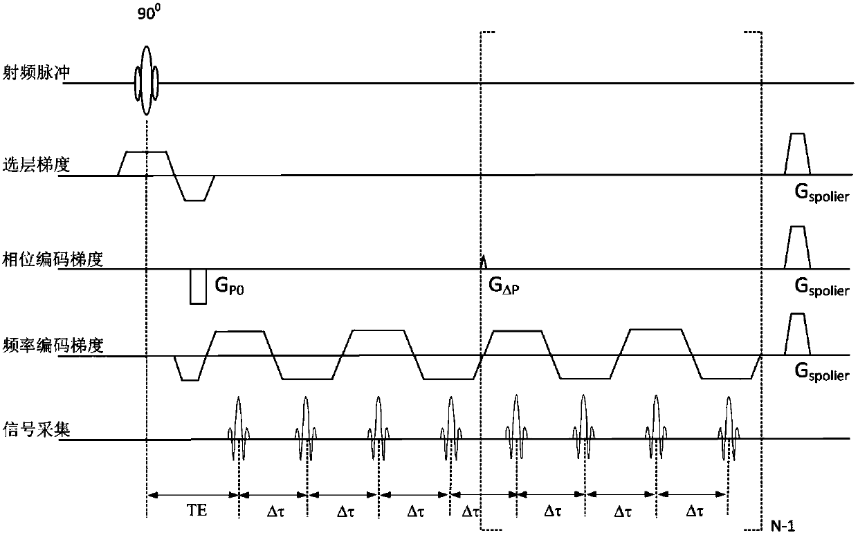
[0056] Loaded on 1.5T MRI system Figure one Show the sequence and set the parameter table, where Dim1=256, Dim2=192, N=4, Δτ=2.3ms, TR=160ms. Execute the sequence and collect 4 groups of T1-weighted echoes, each group consisting of 4 Dixon echoes. The k-space is divided into 4 areas according to the number of echo groups, the area number j is from 1 to 4, and each area is filled with N PE / N=24 k-space lines.

[0057] For the upper half of k-space filling, set the preliminary phase gradient G P0 Is the maximum phase encoding gradient G p 1 / 4, the phase encoding is divided into N PE / N=24 steps, the number of cycles i is from 1 to 24, the first group of echoes fills the central area, and the phase encoding gradient is i·(G P / N) / (N PE / N)=i·G P / 96; the j-th group of echoes fills the j-th area around, the phase encoding gradient is i·G P / 96+(j-1)·G Δp , Where G Δp =G p / 4. For filling the lower half of k-space, set G P0 Just 1 / 4 of the negative maximum value of the phase enc...

Embodiment 2

[0061] Loaded on 3.0T MRI system Figure one Show the sequence and set the parameter table, where Dim1=256, Dim2=192, N=8, Δτ=1.1ms, TR=160ms. Execute the sequence and collect 4 groups of T1-weighted echoes, each group consisting of 4 Dixon echoes. The k-space is divided into 8 regions according to the number of echo groups, the region number j is from 1 to 8, and each region is filled with N PE / N=12 k-space lines.

[0062] For the upper half of k-space filling, set the preliminary phase gradient G P0 Is the maximum phase encoding gradient G P 1 / 8, the phase encoding is divided into N PE / N=12 steps, the number of cycles i is from 1 to 12, the first group of echoes fills the central area, and the phase encoding gradient is i·(G P / N) / (N PE / N)=i·G P / 96; the j-th group of echoes fills the j-th area around, the phase encoding gradient is i·G P / 96+(j-1)·G Δp , Where G Δp =G p / 8. For filling the lower half of k-space, set G P0 Just 1 / 8 of the negative maximum value of the pha...


PUM


Abstract

The invention discloses an ultra-fast single-excitation four-point Dixon water-fat separation imaging method. Single-excitation water-fat separation sequences and data processing modes of multiple versions are adopted, and the method is mainly used for enhancing the signal to noise ratio and increasing the scanning speed to obtain a same-phase diagram, a reverse-phase diagram, a water image and a fat image in which the field asymmetry effect is eliminated and obtain a field distribution diagram delta B0 and a precise distribution diagram (please see the formula in the description). Fast scanning is guaranteed, and meanwhile the clinical diagnosis needs of infants and restless patients are met.

Description

Technical field [0001] The invention relates to the technical field of magnetic resonance imaging, in particular to an ultra-fast single excitation four-point Dixon water-fat separation imaging method. Background technique [0002] The Dixon water-fat separation imaging technology is based on the magnetic resonance gradient echo or spin echo technology, which uses the chemical shift difference of water and fat to perform phase encoding and post-processing the data to obtain the enhanced visualization of the water and fat components in the human anatomical structure. An advanced magnetic resonance imaging technology. The traditional Dixon water-fat separation imaging technology including two-point Dixon and three-point Dixon technology has been widely used in clinical diagnosis, and has unique value for the clear visualization of lesions under fat suppression conditions and the diagnosis of fat diseases. Compared with the two-point Dixon technology, the three-point Dixon technolo...

Claims


Application Information

Patent Timeline
Patent Type & Authority Patents(China)
IPC IPC(8): A61B5/055
CPCA61B5/055
Inventor 罗会俊
Owner 谱影医疗科技(苏州)有限公司
Why Eureka
Try Eureka