Ultrasonic system and multi-image imaging method thereof

An ultrasound system and imaging method technology, applied in ultrasound/sound wave/infrasonic wave diagnosis, sound wave diagnosis, infrasonic wave diagnosis, etc., can solve the problems of inconvenient operation and diagnosis for doctors, smaller size, lower image resolution, etc., to achieve weakened resolution The effect of reducing and improving work efficiency and diagnosis quality

Inactive Publication Date: 2016-10-12
杭州融超科技有限公司
View PDF9 Cites 7 Cited by
  • Summary
  • Abstract
  • Description
  • Claims
  • Application Information

AI Technical Summary

Problems solved by technology

Usually, the display area of ​​the ultrasound image is fixed, such as figure 1 As shown, when the image area is to display multiple images, the size of each image should be reduced accordingly or only part of the image should be displayed; or when the image depth is adjusted deeper, as in Figure 4 As shown, the image is reduced accordingly, which reduces the resolution of the image; these situations will bring inconvenience to the doctor's operation and diagnosis

Method used


Image

  • Ultrasonic system and multi-image imaging method thereof
  • Ultrasonic system and multi-image imaging method thereof
  • Ultrasonic system and multi-image imaging method thereof

Examples


Embodiment

[0028] An ultrasound system such as Figure 7 shown, including the following parts:

[0029] Conventional scanning and processing modules, including transmission, reception, beamforming, mid-processing, digital scan conversion and post-processing;

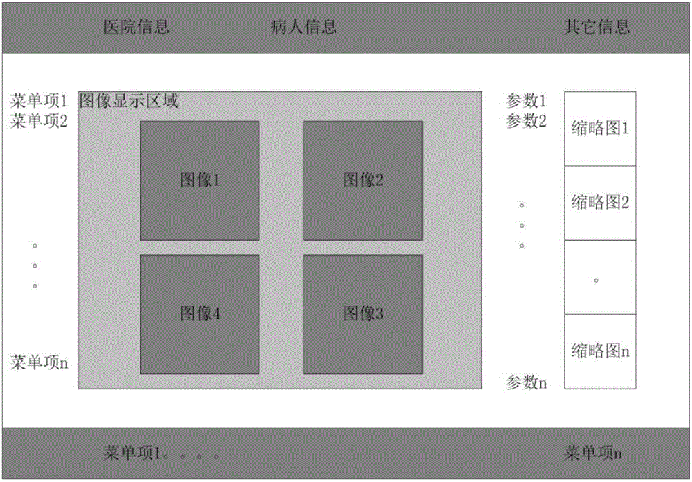
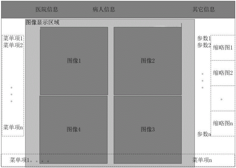
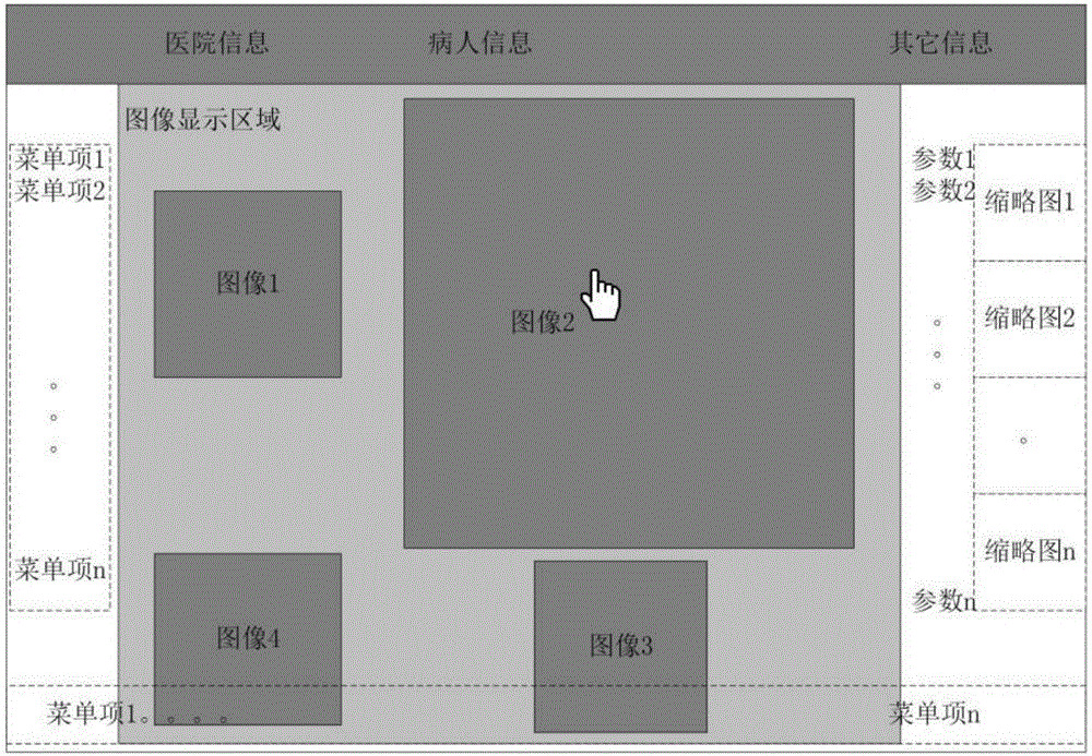
[0030] User interface components, including image display area and non-image display area; the image display area is automatically adjusted or specified by the user depending on the displayed object, and the non-image display area includes image parameters, menu items and other display functional areas, such as hospitals and Patient information area, stored image thumbnail display area.

[0031] in:

[0032] The non-image display area in the user interface component is adjusted or hidden according to the size of the image display area;

[0033] The user interface components include one or more of various control methods such as trackball, stylus, touch and press.

[0034] The multi-image imaging method of the ultrasound system ...


PUM


Abstract

The invention relates to the technical field of ultrasonic imaging, in particular to an ultrasonic system and a multi-image imaging method thereof. The ultrasonic system comprises a conventional scanning and processing module and a user interface assembly. The multi-image imaging method is implemented by adopting range customization of a display area, non-image content automatic concealment, single-image zooming and recovering. According to the method, the size of the display area is increased when the multi-image display or image depth is large, the problem that the resolution is lowered under normal conditions is avoided or reduced, and working efficiency and diagnosis quality of a doctor are improved; particularly, on the portable ultrasonic system, the size of the display screen is limited, the size of a display screen is reasonably utilized, and the method is of great significance in image display.

Description

technical field [0001] The invention relates to the technical field of ultrasonic imaging, in particular to an ultrasonic system and a multi-image imaging method thereof. Background technique [0002] When the ultrasound system is imaging, it supports multiple modes, such as B mode, Color mode, PW, CW mode, M, CM mode, etc. These modes can form dual synchronization modes (such as B / M, B / CM, B / PW, B / CW mode) or tri-synchronous mode (such as B / Color / PW mode), in double-synchronous or tri-synchronous mode, multiple mode images are displayed in the image area at the same time; in order to compare multiple images, the ultrasound system also supports 1 / 2 / 4 multiple image display; in 3D or 4D imaging, the system supports simultaneous display of each section and 3D / 4D images in the image area. Usually, the display area of ​​the ultrasound image is fixed, such as figure 1 As shown, when the image area is to display multiple images, the size of each image should be reduced accordi...

Claims


Application Information

Patent Timeline
Patent Type & Authority Applications(China)
IPC IPC(8): A61B8/00
CPCA61B8/00A61B8/4427A61B8/463A61B8/465A61B8/467A61B8/5207A61B8/5246A61B2560/0431
Inventor 鹿祥鹏
Owner 杭州融超科技有限公司
Why Eureka
Try Eureka