Medical lesion image feature expression method based on region division and Fisher vector

An expression method and image feature technology, applied in the field of medical lesion image feature expression, can solve problems such as limited identification ability, and achieve the effect of strong identification ability and improved accuracy

Inactive Publication Date: 2016-12-07
SOUTHERN MEDICAL UNIVERSITY
View PDF2 Cites 6 Cited by
  • Summary
  • Abstract
  • Description
  • Claims
  • Application Information

AI Technical Summary

Problems solved by technology

Due to the complexity of the texture of the lesion area, the discr

Method used


Image

  • Medical lesion image feature expression method based on region division and Fisher vector
  • Medical lesion image feature expression method based on region division and Fisher vector
  • Medical lesion image feature expression method based on region division and Fisher vector

Examples


experiment example

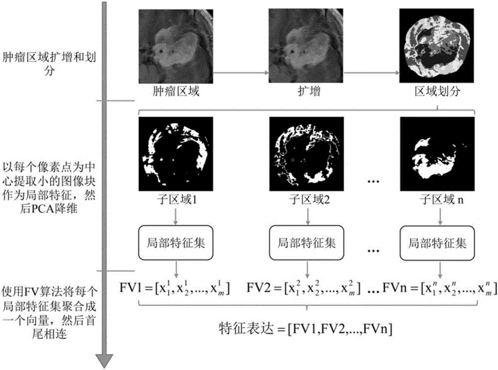
[0048] The data used were T1-weighted contrast-enhanced MRI brain images containing 708 images of meningiomas, 1426 images of gliomas, and 930 images of pituitary tumors. The purpose is to use the feature expression method described in the present invention to retrieve from the database the brain tumor images with the same pathological type as the query image. For the MRI brain tumor images used in the experiment, the selection principle of the parameters is: circular structural elements It is advisable to take the radius of about 24 pixels, the size of the image block to be 9×9, the larger the value of the number N of the region division and the number K of Gaussian in the GMM, the better, but considering the balance between the effect and the calculation efficiency, it is generally taken N=8, K=128.

[0049] In the experiment, 5-fold cross-validation is used to evaluate the experimental results, that is, the data is divided into 5 equal parts, 4 of which are taken as the tra...


PUM


Abstract

The invention relates to the technical field of medical lesion image recognition, in particular to a medical lesion image feature expression method based on region division and a Fisher vector. The method uses an expanded lesion region as a region of interest (ROI), divides the ROI into N sub regions according to the gray values of all pixels in the ROI; in each sub region, extracts small image blocks as local feature descriptors; aggregates the local feature set of each sub region into a vector by using a Fisher vector (FV) algorithm, and then connects the obtained N vectors end to end to obtain the feature expression of a medical lesion image. The feature expression method of the present invention utilizes the region information and space position information around the lesion, and uses the FV algorithm which is more effective than a conventional bag-of-word model to make the constructed feature expression more discriminative, thereby contributing to improving the accuracy of clinical adjuvant diagnosis.

Description

technical field [0001] The invention relates to a feature expression method of medical lesion images based on region division and Fisher vector. Background technique [0002] In modern hospitals, a large number of medical images are generated every day, diagnosed, and then stored in the picture archiving and communication system (PACS). How to effectively use the image data stored in PACS to provide help for clinical decision support, radiologist training, and scientific research in medical schools is a hot spot in the medical field. Content-Based Image Retrieval (Content-Based Image Retrieval, CBIR) is one of the key technologies. The basic idea of ​​CBIR is to construct a suitable feature representation based on the visual content of the image, in order to retrieve images similar to the query image in the database. In the present invention, similar means that the pathological types of the lesions contained in the two images are the same. The retrieval of similar lesions...

Claims


Application Information

Patent Timeline
IPC IPC(8): G06T7/00G06K9/62
CPCG06T7/0012G06T2207/30096G06T2207/10132G06T2207/10081G06T2207/10088G06F18/2132G06F18/23213
Inventor 冯前进陈武凡阳维程君
Owner SOUTHERN MEDICAL UNIVERSITY
Why Eureka
Try Eureka