DNA quantitative analysis method based on cell microscope image

A quantitative analysis and microscope technology, applied in the field of image processing, can solve problems such as time-consuming and missing diploid cells, and achieve high-precision results

Active Publication Date: 2017-01-18
湖南品信生物工程有限公司
View PDF4 Cites 10 Cited by
  • Summary
  • Abstract
  • Description
  • Claims
  • Application Information

AI Technical Summary

Problems solved by technology

However, this type of method will miss a large number of diploid cells when dealing with densely distributed nuclei and large changes in the...

Method used


Image

  • DNA quantitative analysis method based on cell microscope image
  • DNA quantitative analysis method based on cell microscope image
  • DNA quantitative analysis method based on cell microscope image

Examples


Embodiment Construction

[0032] The present invention will be further explained below in conjunction with specific embodiments.

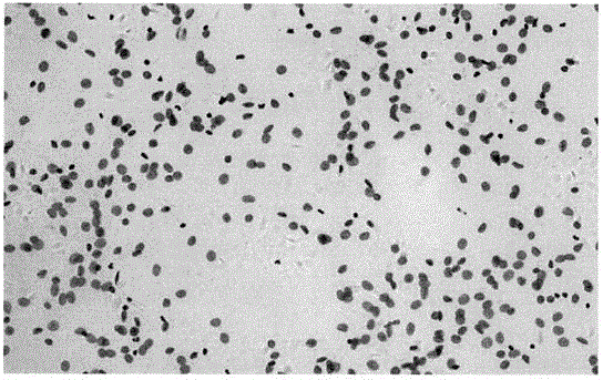
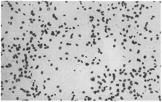
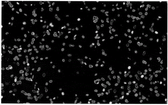
[0033] refer to Figure 1-7 , a kind of DNA quantitative analysis method based on cell microscope image that the present invention proposes, comprises the following steps:

[0034] Step 1: Preprocessing, first perform grayscale processing, extract the r(x, y), g(x, y), b(x, y channels of the original color image, calculate the mean value, and flip it as a grayscale image I(x, y), that is, I(x, y)=(r(x, y)+b(x, y)+g(x, y)) / 3, and then carry out background correction, that is, according to the given Black background BB(x, y) and white background image WB(x, y), perform background correction on image I(x, y) to obtain corrected image I c (x,y):

[0035]

[0036] To further denoise the image, use a Gaussian filter to me c (x, y) for Gaussian smoothing, that is, I s (x, y) = I c (x, y)*h(x, y), in practice, a 5×5 spatial template is used to approximate the Gaussian ...


PUM


Abstract

The invention discloses a DNA quantitative analysis method based on a cell microscope image. The method comprises the following steps that 1) pretreatment is carried out; 2) an MSER algorithm is used to extract candidate areas, and single-tine trees or single nodes are obtained; 3) a simple feature training classifier C1 is extracted from the area on the basis of a training set, the possibility that the classifier belongs to a cell of interest is calculated, and the single-tine trees obtained in the step 2) are further simplified based on the classifier; 4) a statistical, shape and texture feature training classifier C2 are extracted on the basis of simple features of the extraction area of the training set, and the classifier C2 is used to further classify all the candidate areas of positive samples output by the classifier C1; and 5) a DI value of a nucleus is calculated. According to the invention, a maximum stable area algorithm is used to extract a lot of candidate areas from the image, and a cascaded classifier composed of the classifier C1 and the classifier C2 is trained to identify the candidate areas rapidly and accurately.

Description

technical field [0001] The invention relates to the technical field of image processing, in particular to a DNA quantitative analysis method based on cell microscope images. Background technique [0002] Cytological examination is the main way for early screening of typical cancers such as cervical cancer and oral cancer. The smear technique used in the early days was popular because of its low price, but its missed diagnosis rate is high. Although the liquid-based smear technique widely used since the 21st century The effect of film production has been improved, but it requires experienced pathologists to directly observe the morphology of cells under a microscope to give diagnostic results, which is inefficient, easily affected by subjectivity, and has poor repeatability. DNA ploidy analysis technology automatically analyzes the nucleus in the cell image, and then quantitatively measures the chromosome or DNA content in the nucleus, and automatically gives the judgment res...

Claims


Application Information

Patent Timeline
IPC IPC(8): G06T7/00G06T7/11G06K9/62
CPCG06T7/0012G06T2207/10056G06T2207/10024G06T2207/30024G06F18/24323
Inventor 梁毅雄刘剑锋
Owner 湖南品信生物工程有限公司
Why Eureka
Try Eureka