Mammary gland imaging device and control method thereof

A breast imaging and equipment technology, applied in medical science, instruments for radiological diagnosis, diagnosis, etc., can solve problems such as missed diagnosis, subject pain, breast tissue damage, etc., to improve comfort, avoid injury, and improve intelligence The effect of pressure control

Pending Publication Date: 2017-03-08
SHANGHAI UNITED IMAGING HEALTHCARE
View PDF12 Cites 8 Cited by
  • Summary
  • Abstract
  • Description
  • Claims
  • Application Information

AI Technical Summary

Problems solved by technology

In the actual compression process, if the compression force is too small, it may cause uneven compression of the breast, and the image quality does not meet the actual clinical needs, which may lead to missed diagnosis; if the compression force is too large, it will cause pain to the subject, and may even damage the breast. tissue damage
In addition, for different operators, there are also operating errors in different situations, which will also lead to missed diagnosis or discomfort of the examinee.
[0006] To sum up, the current breast imaging equipment will increase the pain of the subject to obtain better quality clinical diagnostic images; or in order to reduce the pain of the subject, the quality of the image can only be sacrificed

Method used


Image

  • Mammary gland imaging device and control method thereof
  • Mammary gland imaging device and control method thereof
  • Mammary gland imaging device and control method thereof

Examples


Embodiment 1

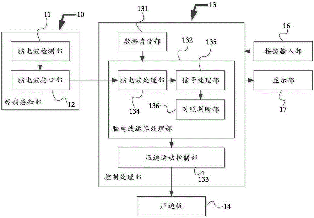
[0037] figure 2 It is a schematic structural frame diagram of a breast imaging device according to the first embodiment of the present invention. Such as figure 2 As shown, the breast imaging device includes: a compression board 14, which compresses the breast; a control processing unit 13, which controls the compression board 14 to compress the breast; also includes a pain sensing unit 10, which is used to sense the physiological pain of the subject information, and send the physiological pain information to the control processing unit 13; the control processing unit 13 is further configured to control the compression plate to compress the mammary gland according to the physiological pain information.

[0038] Here, the pain sensing unit 10 senses by analyzing brain waves. In this embodiment, the pain sensing unit 10 is an electroencephalogram sensor as an example for illustration. Specifically, the pain sensing unit 10 includes: an EEG detection unit 11 and an EEG interf...

Embodiment 2

[0044] Figure 4 It is a schematic structural frame diagram of a breast imaging device according to the second embodiment of the present invention. Such as Figure 4 As shown, the breast imaging device includes: a compression board 24, which compresses the breast; a control processing unit 23, which controls the compression board 24 to compress the breast; and also includes a pain sensing unit 20, which is used to sense the subject’s physiological pain information, and send the physiological pain information to the control processing unit 23; the control processing unit 23 is also used to control the compression plate to compress the mammary gland according to the physiological pain information; the pressure detection unit 28 is used to Sensing the compression force of the compression plate 24 on the mammary gland; the fine-tuning part 25 fine-tunes the compression plate 24 in a small range.

[0045] The pain sensing unit 20 is the same as that in Embodiment 1, and will not ...


PUM


Abstract

The invention discloses a mammary gland imaging device. The device comprises a compressing plate used for compressing the mammary gland, and a control processing part used for controlling the compressing plate to compress the mammary gland, and further comprises a pain perception part used for perceiving physiologic pain information of an examinee and sending the pain information to the control processing part, wherein the control processing part is further used for controlling the compressing plate to compress the mammary gland according to the physiologic pain information. According to the mammary gland imaging device, the compressing operation of the mammary gland compressing plate is controlled by detecting the pain information of the examinee; the compression is continuously applied when a compressing upper limit value is not exceeded, and the pain information of the examinee is detected; and once the examinee is discovered to feel uncomfortable or painful, the situation can be fed back to the control processing part through the pain perception part, and the compression on the mammary gland is stopped, so that the injury caused by the fact that the examinee feels painful or the mammary gland is excessively oppressed can be avoided and a relatively user-friendly and intelligent control effect is achieved.

Description

technical field [0001] The invention relates to the field of medical equipment, in particular to breast imaging equipment for controlling the compression force of a compression plate according to the detected physiological pain information of a subject. Background technique [0002] With the development of computer science and information technology, medical imaging technology has also been developed rapidly, and various medical imaging systems are emerging. Mammography (Mammography) equipment and digital breast tomosynthesis (DBT, digital breast tomosynthsis) equipment have been widely used as breast cancer screening and diagnosis. [0003] figure 1 is a schematic diagram of an existing breast imaging device. Such as figure 1 As shown, the breast imaging device 1 includes: a column 2 , a rotating arm 3 , a tube 4 , a compression plate 5 and a casing 6 . The frame of the mammography equipment generally includes a column 2 and a rotating arm 3 . The bulb 4 is an X-ray so...

Claims


Application Information

Patent Timeline
Patent Type & Authority Applications(China)
IPC IPC(8): A61B6/00
Inventor 张娜
Owner SHANGHAI UNITED IMAGING HEALTHCARE
Why Eureka
Try Eureka