
Mammary gland ultrasonic imaging device and probe scanning mechanism

A technology of breast imaging and scanning mechanism, which is applied in mammography, structure of ultrasound/sound wave/infrasound diagnostic equipment, ultrasound/sound wave/infrasound image/data processing, etc. Impairment of imaging quality and other issues, to achieve the effect of simple structure, reduced deformation, and accurate scanning position

Pending Publication Date: 2017-03-22
CHISON MEDICAL TECH CO LTD
View PDF17 Cites 4 Cited by
  • Summary
  • Abstract
  • Description
  • Claims
  • Application Information

AI Technical Summary

Problems solved by technology

[0002] At present, the application of ultrasonic diagnostic instruments in clinical diagnosis has been very popular, and it has made great contributions to doctors' accurate understanding of patients' conditions and formulation of medical plans. However, the application in physical examination is still insufficient, especially in the screening of breast diseases. During the screening, due to the requirements of screening time and screening intensity, manual touch screening is still the main method, supplemented by ultrasound scanning confirmation, which cannot guarantee the accuracy of screening well, and the labor intensity of doctors is also very high.
Existing ultrasonic mammary gland examination is supine, at this time the mammary gland is easily compressed by the transducer, which has a certain impact on the imaging quality of the image
In addition, the existing mammary gland detection device can only perform ultrasound examination during detection, and has no real-scene camera function for mammary glands. When the doctor wants to observe the physical picture of the mammary gland, it cannot be traced back or diagnosed on the spot

Method used


Image

  • Mammary gland ultrasonic imaging device and probe scanning mechanism
  • Mammary gland ultrasonic imaging device and probe scanning mechanism
  • Mammary gland ultrasonic imaging device and probe scanning mechanism

Examples


Embodiment Construction

[0027] The present invention will be further described below in conjunction with specific drawings.

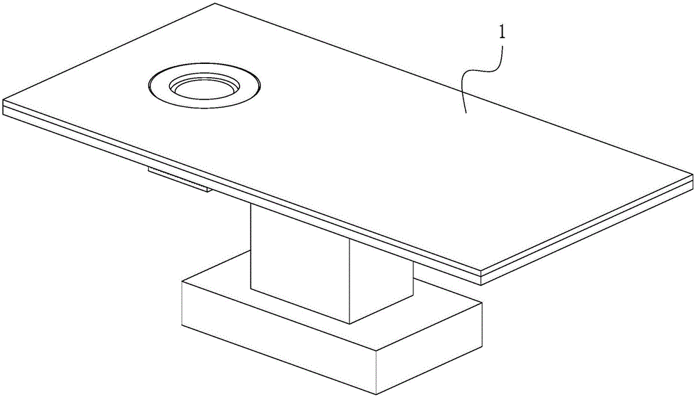
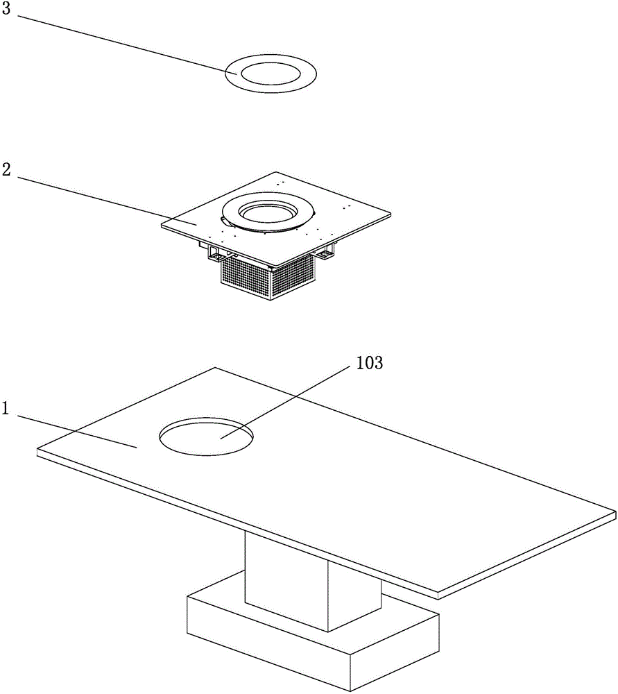
[0028] Such as figure 1 , figure 2 As shown, the breast imaging device of the present invention includes a detection bed 1 and a probe scanning mechanism 2; an opening 103 is provided on the detection bed 1, and the probe scanning mechanism 2 is installed in the opening 103, and the end surface of the probe scanning mechanism 2 Install cushion 3 on it.

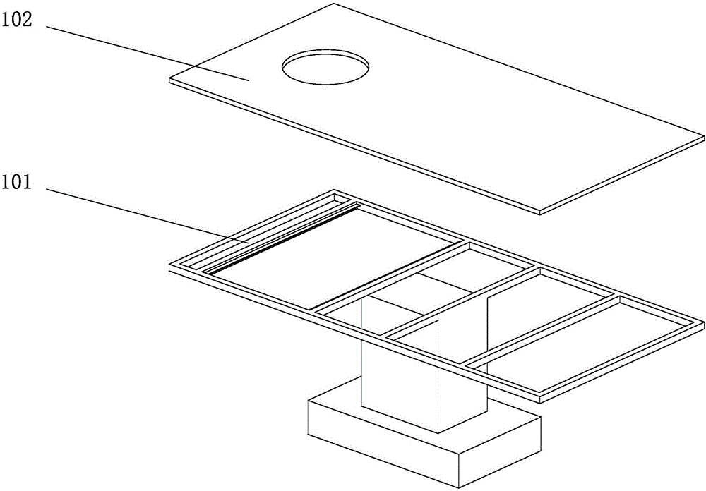
[0029] Such as Figure 3-1 As shown, the test bed 1 includes a liftable bed frame 101 and a bed board 102 , and the bed board 102 is installed on the liftable support 101 . Such as Figure 3-2 As shown, the bed board 102 includes a wooden board 102a, sponges 102b, 1 and soft leather 102c, and the wooden board 102a, sponge 102b and soft leather 102c are installed sequentially from bottom to top.

[0030] Such as Figure 4 , Figure 5 As shown, the probe scanning mechanism 2 includes a rotating mechanism 200 , an electri...


PUM


Abstract

The invention relates to a mammary gland ultrasonic imaging device which comprises a detection bed and a probe scanning mechanism, wherein an open hole for mounting the probe scanning mechanism is formed in the detection bed. The mammary gland ultrasonic imaging device is characterized in that the probe scanning mechanism comprises a rotary mechanism and a camera component, wherein the rotary mechanism comprises a cup body, a cup holder, a transducer component and a drive device; the cup body is provided with an accommodating cavity; the cup body and the cup holder are fixed on a mounting plate; the transducer component is mounted in the cup holder; the transducer component is fixedly connected with one-end output rotation shaft of the drive device; the one-end output rotation shaft of the drive device is rotatably connected with the cup holder; the other-end output rotation shaft of the drive device is a rotation condition detection end; the transducer component is connected with one end of a cable, and the other end of the cable is connected with a transfer end of a probe through the cup holder; and one or more camera components are mounted on the lateral part of the rotary mechanism. The device provided by the invention can realize no-extrusion or no-compression detection as well as automatic photographing or image pickup.

Description

technical field [0001] The invention relates to a breast ultrasound imaging device, which belongs to the technical field of ultrasound imaging devices. Background technique [0002] At present, the application of ultrasonic diagnostic instruments in clinical diagnosis has been very popular, and it has made great contributions to doctors' accurate understanding of patients' conditions and formulation of medical plans. However, the application in physical examination is still insufficient, especially in the screening of breast diseases. During the screening, due to the screening time and screening intensity requirements, manual touch screening is still the main method, supplemented by ultrasound scanning confirmation, which cannot guarantee the accuracy of screening, and the labor intensity of doctors is also very high. The existing ultrasonic mammary gland examination is performed in a supine position. At this time, the mammary gland is easily compressed by the transducer, wh...

Claims


Application Information

Patent Timeline
Patent Type & Authority Applications(China)
IPC IPC(8): A61B8/08
CPCA61B8/0825A61B8/40A61B8/44A61B8/4444A61B8/4483
Inventor 韩旭宫明晶高峰孙冬冬欧阳齐钦赵明昌
Owner CHISON MEDICAL TECH CO LTD
Learn More