
VR operation simulation system based on human organ 3D model and method

A technology for surgical simulation and human organs, applied in the field of medical image processing, can solve the problems of inability to achieve preoperative simulation resection and surgical risk assessment, and inability to achieve organ segmentation, so as to improve surgical skills and proficiency, increase authenticity, Difficulty-reducing effect

Inactive Publication Date: 2017-06-09
厦门强本宇康科技有限公司
View PDF8 Cites 10 Cited by
  • Summary
  • Abstract
  • Description
  • Claims
  • Application Information

AI Technical Summary

Problems solved by technology

However, for the current 3D visualization technology of medical images, the segmentation of organs cannot be realized, and it is difficult for doctors to intuitively see which segment the tumor is in from the 3D image
In addition, the current 3D visualization technology of medical images only provides 3D viewing function, but cannot realize preoperative simulated resection and surgical risk assessment

Method used


Image

  • VR operation simulation system based on human organ 3D model and method

Examples


Embodiment 1

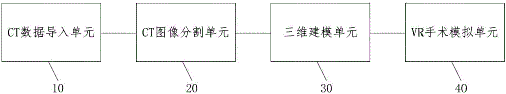
[0042] refer to figure 1 As shown, the present invention discloses a VR surgery simulation system based on a 3D model of human organs, including a CT data import unit 10, a CT image segmentation unit 20, a three-dimensional modeling unit 30 and a VR surgery simulation unit 40, wherein:

[0043] The CT data import unit 10 is used for importing CT data, and the CT data includes CT images, patient information, hospital information, CT machine model information and CT slice accuracy information. The CT data importing unit 10 has a CT data preview module and a CT image preprocessing module, the CT data preview module is used to view relevant content of the CT data, and the CT image preprocessing module is used to judge whether the CT image is inverted, and the inverted CT image Adjust to the correct orientation.

[0044] The CT image segmentation unit 20 is used to segment soft tissue organs, tumors, blood vessels and hard tissues on the CT image, which includes a soft tissue orga...

Embodiment 2

[0048] The present invention discloses a VR surgery simulation method based on a 3D model of human organs, which is implemented based on the VR surgery simulation system of Embodiment 1. The method includes the following steps:

[0049] S1. Using the CT data import unit 10 to import CT data.

[0050] S2. Using the CT image segmentation unit 20 to segment soft tissue organs, tumors, blood vessels and hard tissues on the CT image, and segment the soft tissue organs according to the blood vessels. This step is achieved through the following sub-steps:

[0051] S21. Segmentation of soft tissue organs, selecting positions of soft tissue organs on the CT image through human-computer interaction, and automatically segmenting soft tissue organs. The position of the soft tissue organ on the CT image is selected by placing seed points through human-computer interaction, and the computer automatically segments the soft tissue organ. For large soft tissue organs, the position of the soft...


PUM


Abstract

The invention discloses a VR operation simulation system based on a human organ 3D model. The system comprises a CT data importing unit, a CT image segmentation unit, a 3D modeling unit and a VR operation simulation unit, wherein the CT data importing unit is used for importing the CT data; the CT image segmentation unit is used for segmenting soft tissue organs, tumors, blood vessels and hard tissues on the CT image and sectioning the soft tissue organs according to the blood vessels; the 3D modeling unit is used for establishing the 3D models of soft tissue organs, tumors, blood vessels and hard tissues in all sections and fusing to form the human organ 3D model; the VR operation simulation unit comprises a VR operation simulation excision module, an excision result analysis module, an operation simulation measuring module and an operation simulating risk assessment module. The invention also discloses a VR operation simulation method based on the human organ 3D model. According to the invention, the section of the organ with the tumor is directly acquired and the positioning for the tumor is more accurate, so that the operation difficulty and risk are reduced; the preoperative simulation excision and operation risk assessment can be realized; a virtual reality technology is adopted for simulating the operation; the authenticity of the analog simulation and the accuracy of the simulation result are increased.

Description

technical field [0001] The invention relates to the technical field of medical image processing, in particular to a VR surgery simulation system and method based on a 3D model of human organs. Background technique [0002] With the development of science and technology, human beings have made a lot of progress in medicine, but in clinical medicine, there are still problems of low surgical success rate and high risk for some diseases. [0003] At present, for operations on soft tissue organs such as the liver and lungs, since the tumor is usually inside the organ, doctors still need to find the location of the tumor based on experience during the operation. The main problem is that it is difficult for the doctor to find the location of the tumor. It also increases the risk of surgery. As we all know, some organs in the human body are segmented organs. For example, the liver can be divided into five lobes and eight segments through the arteries, and the lungs can be divided i...

Claims


Application Information

Patent Timeline
IPC IPC(8): A61B34/10
Inventor 王博亮
Owner 厦门强本宇康科技有限公司
Learn More