
Method for measuring related parameters of radiation dosage in X-ray tomosynthesis

A method for measuring and related parameters, used in the medical field

Inactive Publication Date: 2017-06-20
NANFANG HOSPITAL OF SOUTHERN MEDICAL UNIV
View PDF0 Cites 5 Cited by
  • Summary
  • Abstract
  • Description
  • Claims
  • Application Information

AI Technical Summary

Problems solved by technology

But there is no simple method to calculate the product of air kerma and dose area

Method used


Image

  • Method for measuring related parameters of radiation dosage in X-ray tomosynthesis
  • Method for measuring related parameters of radiation dosage in X-ray tomosynthesis
  • Method for measuring related parameters of radiation dosage in X-ray tomosynthesis

Examples


Embodiment Construction

[0018] The following will clearly and completely describe the technical solutions in the embodiments of the present invention with reference to the accompanying drawings in the embodiments of the present invention. Obviously, the described embodiments are only some, not all, embodiments of the present invention. Based on the embodiments of the present invention, all other embodiments obtained by persons of ordinary skill in the art without making creative efforts belong to the protection scope of the present invention.

[0019] Digital tomographic fusion and post-processing technology is an important supplement and extension method of conventional X-ray examination, and has unique advantages in the diagnosis of lesions in deep parts and complex parts of anatomy. In the past ten years, with the improvement of computer processing capabilities and the successful development of digital flat panel detectors, digital tomographic fusion technology and digital reconstruction have emerg...


PUM


Abstract

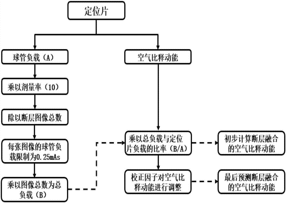
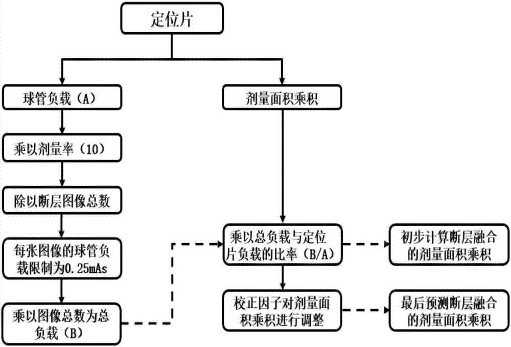
The invention discloses a method for measuring radiation dose-related parameters of X-ray tomographic fusion. The method of the present invention includes: according to the characteristics of the X-ray tomographic fusion system, the tube load of the first positioning film is multiplied by the dose rate to obtain the total load of the system, and then evenly distributed to each tomographic image. According to the rounding principle of the Renard sequence, the tube load of each tomographic image was limited to a minimum of 0.25 mAs, and multiplied by the total number of exposures to obtain the total tube load of the entire tomographic fusion. The total tube load was divided by the tube load of the spacer, and the obtained ratio was multiplied by the product of air kerma and dose area of ​​the spacer, and the product of air kerma and dose area of ​​tomosynthesis was preliminarily obtained. The parameters are adjusted by two correction factors to obtain the final predicted air kerma and dose-area product. The method of the present invention is simple and effective, and does not need to separately superimpose the exposure parameters of all different layers.

Description

technical field [0001] The invention belongs to the field of medical technology, and relates to a simple method for calculating X-ray radiation dose, in particular to a method for measuring radiation dose-related parameters of X-ray tomographic fusion. Background technique [0002] In 1895, German physicist Roentgen discovered X-rays and successfully took X-ray images of his wife's palm in the same year. Since 1897, X-ray imaging technology has been formally used clinically to perform plain film shooting, fluoroscopy and tomosynthesis (Digital Tomosynthesis, DTS) for patients. For example, in chest imaging, X-ray detection of the lungs is of great significance to the early detection of lung cancer; fractures are the most common disease in emergency examinations, and plain X-ray films can make a rapid and accurate diagnosis of fracture locations ; For total gastrointestinal radiography, contrast agents can clearly show the location of gastrointestinal lesions under X-ray flu...

Claims


Application Information

Patent Timeline
IPC IPC(8): A61B6/00A61B6/03
CPCA61B6/52A61B6/03
Inventor 马乐蔡裕兴秦耿耿陈卫国
Owner NANFANG HOSPITAL OF SOUTHERN MEDICAL UNIV
Learn More