Nasopharyngeal carcinoma primary lesion clinical target area automatic drawing method based on mutual correlation rule

A technology for primary disease and nasopharyngeal carcinoma, which is applied in the research field of simulating tumor spread to achieve the effect of improving work efficiency

Active Publication Date: 2017-06-20
PERCEPTION VISION MEDICAL TECH CO LTD
View PDF3 Cites 12 Cited by
  • Summary
  • Abstract
  • Description
  • Claims
  • Application Information

AI Technical Summary

Problems solved by technology

The existing ABAS target area delineation system can only be implemented in the three-dimensional direction, and the numerical range specified by the user is rigidly expanded as the preventive irradiation target area

Method used


Image

  • Nasopharyngeal carcinoma primary lesion clinical target area automatic drawing method based on mutual correlation rule
  • Nasopharyngeal carcinoma primary lesion clinical target area automatic drawing method based on mutual correlation rule
  • Nasopharyngeal carcinoma primary lesion clinical target area automatic drawing method based on mutual correlation rule

Examples


Embodiment

[0047] In this embodiment, a method for automatically delineating clinical target volumes of primary lesions of nasopharyngeal carcinoma based on correlation rules comprises the following steps:

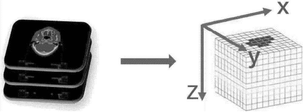
[0048] S1. Generate a binary image of the nasopharyngeal primary tumor area, such as figure 1 As shown, the specific steps of binary image generation are:

[0049] S101. Read the tumor outline file drawn by the doctor, set the image pixel grid corresponding to the tumor area to 1, and set the pixel grid of the tumor-free area to 0.

[0050] The tumor area will use the method of judging whether the pixel points are within the polygon (InPolygon) to separate the non-tumor area and the tumor area. The specific calculation formula of InPolygon is:

[0051] IN=InPolygon(x,y,xv,yv)

[0052] In the formula, IN is the output binary image, and its size is the same as the original input CT image I(x, y), (x, y) is the coordinate of the pixel point, and (xv, yv) is the point coordinate on th...


PUM


Abstract

The invention discloses a nasopharyngeal carcinoma primary lesion clinical target area automatic drawing method based on a mutual correlation rule. The method comprises a step of generating a binary image of a nasopharyngeal primary tumor area, a step of removing instable data of a tumor outermost layer, a step of carrying out data discretization operation on the image sequence of a template CT, and dividing the image sequence and generating corresponding three-dimensional grids, a step of counting the frequency that voxel combination appears, and establishing a database of a correlation rule, a step of reading new patient gross tumor data with the need of drawing a clinical target area, using the mutual correlation rule, simulating tumor growth, and automatically drawing a clinical radiotherapy target area. Compared with manual drawing at present, the automation of drawing is realized by the system, and the working efficiency of a doctor is improved. A drawing result reaches a drawing standard required by the doctor, and local adjustment is taken as post processing and is provided for the doctor for selection.

Description

technical field [0001] The invention relates to the research field of simulating tumor diffusion, in particular to a method for automatically delineating clinical target areas of nasopharyngeal carcinoma primary lesions based on mutual correlation rules. Background technique [0002] Intensity Modulated Radiation Therapy (IMRT) is currently the mainstream radiotherapy technique for the treatment of nasopharyngeal carcinoma. Accurate target delineation is the prerequisite for successful clinical radiotherapy planning. The quality of life of patients after treatment has a very critical impact. At present, the delineation of the target area mainly relies on the manual delineation of doctors. The ABAS target area delineation system currently used in the hospital realizes the rigid expansion of the numerical range specified by the doctor as the preventive irradiation target area in the three-dimensional direction, and then, according to the clinical experience of the doctor, com...

Claims


Application Information

Patent Timeline
Patent Type & Authority Applications(China)
IPC IPC(8): G06T5/00G06T15/00G06T7/11
CPCG06T5/005G06T5/009G06T15/005G06T2207/30096G06T2207/10088
Inventor 陆遥孙颖喻莎田皎林丽李赋
Owner PERCEPTION VISION MEDICAL TECH CO LTD
Why Eureka
Try Eureka