
A method and kit for isolating high-purity urinary exosomes

An exosome, high-purity technology, applied in the field of separating high-purity urine exosomes, can solve the problems of low exosome purity, expensive kits, and insufficient exosome purity.

Active Publication Date: 2021-05-18
SHANGHAI JIAO TONG UNIV
View PDF3 Cites 0 Cited by
  • Summary
  • Abstract
  • Description
  • Claims
  • Application Information

AI Technical Summary

Problems solved by technology

But the purity of exosomes obtained is not high (Danqi Wang and Wei Sun, Proteomics, 2014; 14, 1922-1932)
[0006] The third method is a commercial kit. This method can be divided into two categories. One is to add the extraction reagent to the sample and centrifuge to obtain the exosome precipitation (Lobb RJ et al. J Extracell Vesicles2015; 4:27031), but the shortcomings of this method are that the extracted exosomes are not pure enough and the kits are expensive
[0007] The fourth method is to use cheap polyethylene glycol to precipitate exosomes (Rider MA et al. SciRep 2016; 6:23978), which is similar to the principle of commercial exosome extraction kits. The advantage is that the extraction cost is not high, and the disadvantage is that the purity of the extracted exosomes is not high

Method used


Image

  • A method and kit for isolating high-purity urinary exosomes
  • A method and kit for isolating high-purity urinary exosomes
  • A method and kit for isolating high-purity urinary exosomes

Examples


Embodiment 1

[0041] 1. Centrifuge the freshly obtained urine at 3000rpm for 5 minutes. The precipitate is the cells in the urine. Remove the precipitate and take the supernatant.

[0042] 2. Filter the supernatant obtained in step 1 through a 0.22 micron filter head to remove larger impurities.

[0043] 3. Take 10 mL of the filtrate obtained in step 2 and add it into a 300 kDa dialysis bag (Spectrum, USA). Dialyze in PBS for 9 hours, replace PBS every 3 hours, and use a total of 2 L of PBS. Miscellaneous proteins and other molecules are dialyzed out, leaving exosomes behind.

[0044] 4. Add the exosomes in step 3 into a 100kDa ultrafiltration tube, and perform ultrafiltration at 3000rpm for 5min to concentrate the volume of exosomes.

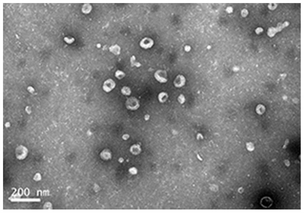
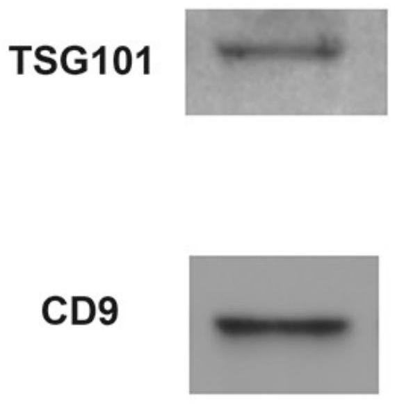
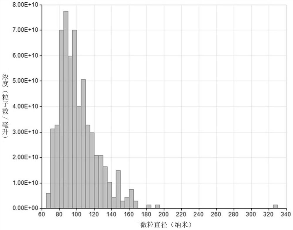
[0045] The present invention uses transmission electron microscopy (TEM), Western Blot, and qNano to characterize the obtained urine exosomes, and the specific characterization results are as follows:

[0046] (1) Transmission electron microscope (TEM) ph...

Embodiment 2

[0054] 1. Centrifuge the freshly obtained urine at 3000rpm for 5min. The precipitate is the cells in the urine. Remove the precipitate and take the supernatant.

[0055] 2. Filter the supernatant obtained in step 1 through a 0.22 micron filter head to remove larger impurities.

[0056] 3. Collect the filtrate obtained in step 2 and store it in a freezer at -80°C for 3 days.

[0057] 4. Thaw the urine frozen in step 3 at room temperature

[0058] 5. Add 10 mL of the melted urine obtained in step 4 into a 300 kDa dialysis bag. Dialyze in 2L PBS for 9 hours, and change PBS every 3 hours. Miscellaneous proteins and other molecules are dialyzed out, leaving exosomes behind.

[0059] 6. Add the exosomes in step 5 into a 100kDa ultrafiltration tube, and perform ultrafiltration at 3000rpm for 5min to concentrate the volume of exosomes.

Embodiment 3

[0061] 1. Centrifuge the freshly obtained urine at 3000rpm for 5min. The precipitate is the cells in the urine. Remove the precipitate and take the supernatant.

[0062] 2. Filter the supernatant obtained in step 1 through a 0.22 micron filter head to remove larger impurities.

[0063] 3. Take 10 mL of the filtrate obtained in step 2 and add it to a 300 kDa dialysis bag. Dialyze in 2L 0.9% normal saline for 9 hours, and change 0.9% normal saline every 3 hours. Miscellaneous proteins and other molecules are dialyzed out, leaving exosomes behind.

[0064] 4. Add the exosomes in step 3 into a 100kDa ultrafiltration tube, and perform ultrafiltration at 3000rpm for 5min to concentrate the volume of exosomes.

[0065] For Examples 2 and 3, the present invention uses the present invention to characterize the obtained exosomes using a transmission electron microscope (TEM), Western Blot, and qNano. The detection results are similar and will not be repeated here.

[0066] The prese...


PUM

PropertyMeasurementUnit
pore sizeaaaaaaaaaa
diameteraaaaaaaaaa
particle sizeaaaaaaaaaa

Abstract

The invention provides a method for separating high-purity urine exosomes, which is characterized in that it comprises the following steps: step 1, centrifuging the urine sample, discarding the precipitate, and collecting the supernatant; step 2, filtering the supernatant with a filter head, collecting Filtrate; step 3, put the filtrate obtained in step 2 into a dialysis bag for dialysis to obtain exosomes; step 4, ultrafilter the exosomes obtained in step 3 in an ultrafiltration tube to finally obtain concentrated urine exosomes secretion body. The invention also provides a kit for isolating high-purity urinary exosomes. Compared with the current conventional urine exosome isolation method, the present invention has the advantages of low cost, simple operation, high yield and purity, and complete exosome structure.

Description

technical field [0001] The invention belongs to the field of biotechnology, and in particular relates to a method and a kit for isolating high-purity urinary exosomes. Background technique [0002] Exosomes are membrane vesicles with a diameter of about 30-150nm that can be secreted by many cells and contain a variety of proteins and RNAs. They have the function of transmitting information between cells. Different cells may secrete specific proteins and RNAs. Studies have shown that urine also contains exosomes. [0003] One of the difficulties in the study of urinary exosomes is the extraction and separation of exosomes in urine. The existing separation methods for separating exosomes mainly include the following: [0004] The first approach is to use different ultracentrifugation speeds (Théry C et al. Curr Protoc CellBiol, 2006; 3:1–29) or density gradient centrifugation (Tauro BJ et al. Methods 2012; 56:293–304.) , this method is considered to be the gold standard fo...

Claims


Application Information

Patent Timeline
Patent Type & Authority Patents(China)
IPC IPC(8): C12N5/071
CPCC12N5/0684G01N2333/435
Inventor 崔大祥杨蒙郅晓刘岩磊
Owner SHANGHAI JIAO TONG UNIV
Learn More