
Deep-convolution-neural-network-based CT pulmonary nodule detection method

A neural network and deep convolution technology, applied in the field of CT pulmonary nodule detection based on deep convolutional neural network, can solve the problems of shallow network structure and failure to make full use of deep learning, etc., achieve strong adaptability and improve prediction ability , high efficiency effect

Active Publication Date: 2017-08-04
ZHEJIANG UNIV
View PDF9 Cites 179 Cited by
  • Summary
  • Abstract
  • Description
  • Claims
  • Application Information

AI Technical Summary

Problems solved by technology

However, the network structure designed by this method is relatively shallow, with only three convolutional layers, and it is necessary to train network models of multiple scales for fusion
And this method still uses the traditional method to recommend candidate nodules, which fails to take full advantage of the advantages of deep learning

Method used


Image

  • Deep-convolution-neural-network-based CT pulmonary nodule detection method
  • Deep-convolution-neural-network-based CT pulmonary nodule detection method
  • Deep-convolution-neural-network-based CT pulmonary nodule detection method

Examples


Embodiment Construction

[0042] The specific implementation manners of the present invention will be further described in detail below in conjunction with the accompanying drawings and embodiments. The following examples are used to illustrate the present invention, but are not intended to limit the scope of the invention:

[0043] The data used in the specific experiments of the present invention come from the LIDC / IDRI database, including CT images of 888 patients, including 1086 nodules in total. The data was obtained by four experienced chest radiologists who performed two image annotations on the patient's CT images. The first time was blind labeling, and the second time was to refer to other doctors to correct the results. However, these 888 CT images are collected by different instruments, and the pixel intervals of CT images are different, and the range of variation is large. 0.46-0.97mm. At the same time, the HU range and contrast of these CT images are also different.

[0044] Such as fi...


PUM


Abstract

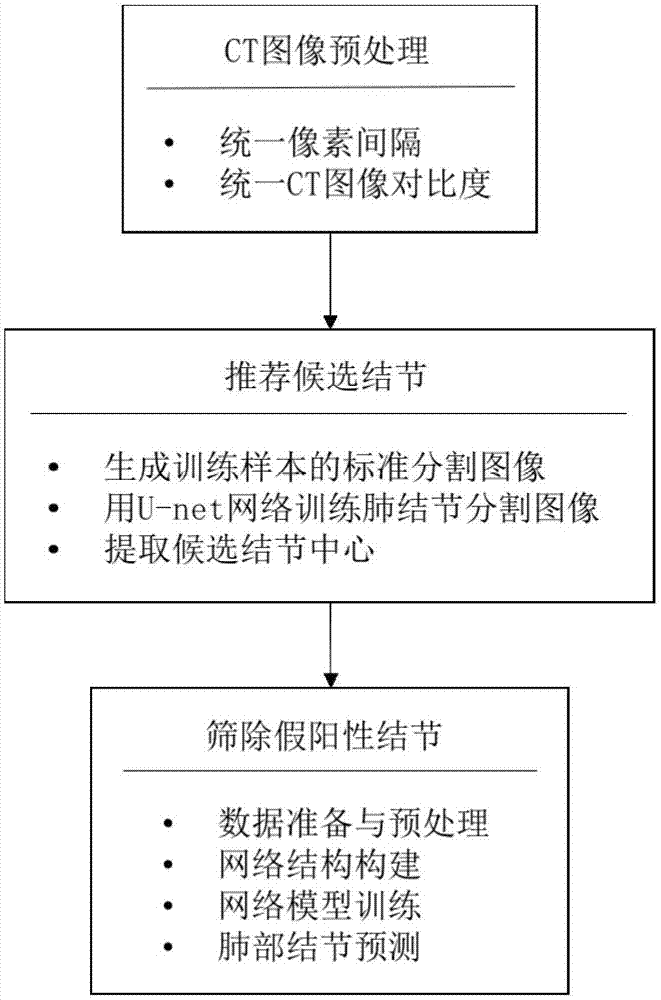
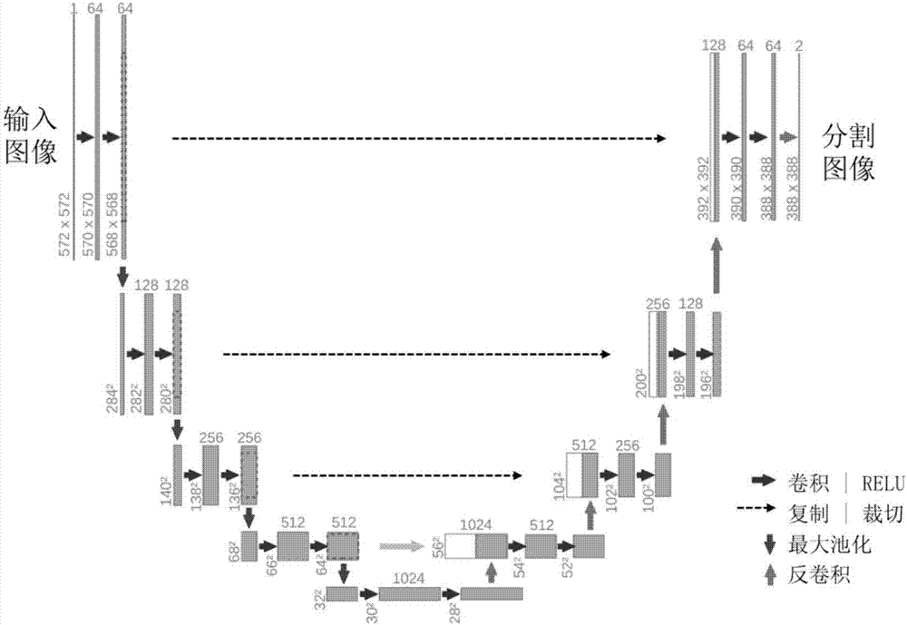
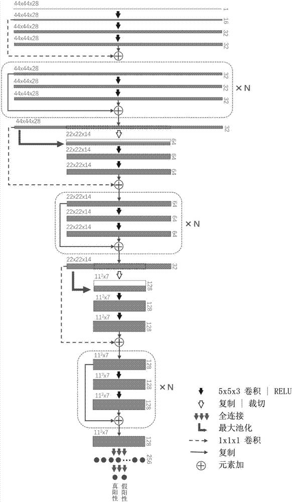
The invention discloses a deep-convolution-neural-network-based CT pulmonary nodule detection method. The method comprises: (1), CT image pretreatment is carried out, so that the pixel spacing becomes unified and image comparison is unified; (2), a two-dimensional convolution neural network U-net is trained, a pulmonary nodule segmentation image is predicted, and a candidate nodule is recommended based on the pulmonary nodule segmentation image; and (3), a three-dimensional deep residual neural network Resnet3D is trained, a true-false positive probability of the pulmonary nodule is predicted, and a false positive nodule is screened out. According to the detection method disclosed by the invention, the deep learning advantages are utilized fully, so that the pulmonary nodule can be detected in a CT image automatically, efficiently and accurately; and the adaptability to medical big data is high.

Description

technical field [0001] The present invention relates to a method for detecting pulmonary nodules aimed at CT images, in particular to a method for detecting pulmonary nodules in CT based on a deep convolutional neural network. Background technique [0002] Lung cancer is the main cause of cancer-related deaths worldwide. Using CT scans to check high-risk groups is an effective means of detecting early lung cancer. However, the number of such groups is huge, and the workload of radiologists has increased dramatically. Therefore, computer-assisted Diagnosis plays a very important role. [0003] At present, in the field of computer-aided detection of pulmonary nodules in CT images, a lot of research work has been carried out based on traditional statistical machine learning methods, and some results have been achieved. The detection step is usually divided into two steps. The first step is to recommend candidate nodules and detect areas where nodules may exist in the lung CT i...

Claims


Application Information

Patent Timeline
IPC IPC(8): G06T7/00G06T7/10G06N3/04G06N3/08
CPCG06T7/0012G06T2207/10081G06T2207/20081G06T2207/20084G06T2207/30064
Inventor 金弘晟李宗曜童若锋
Owner ZHEJIANG UNIV
Learn More