Dual-channel structure optical digital phase contrast microscopy imaging system and realization method thereof

A technology for microscopic imaging and implementation methods, applied in microscopes, optics, optical components, etc., can solve the problems of refresh speed limiting image acquisition frame rate, inability to apply phase contrast imaging, affecting modulation effects, etc., to solve the problem of expensive devices, Achieve high-speed digital phase contrast imaging with less external interference

Pending Publication Date: 2017-08-08
GUANGDONG OPTO MEDIC TECH CO LTD
View PDF5 Cites 17 Cited by
  • Summary
  • Abstract
  • Description
  • Claims
  • Application Information

AI Technical Summary

Problems solved by technology

However, according to the introduction in the literature, this method uses a spatial light modulator to modulate the light in the spectral plane of the imaging optical path, which is expensive, and the refresh rate of the spatial light modulator limits the frame rat

Method used


Image

  • Dual-channel structure optical digital phase contrast microscopy imaging system and realization method thereof
  • Dual-channel structure optical digital phase contrast microscopy imaging system and realization method thereof
  • Dual-channel structure optical digital phase contrast microscopy imaging system and realization method thereof

Examples


Embodiment 1

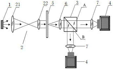
[0030] like figure 1 As shown, this embodiment relates to a dual-channel structured light digital phase-contrast microscopic imaging system, including a light source 1, a beam expander collimator unit 2, a spectroscopic device 3, a lens group and two identical cameras 4, specifically, the The light source 1 is preferably an LED light source with a luminous spectral bandwidth of about 20nm. The beam expander and collimation unit 2 is composed of a first lens 21 and a second lens 22. The lens group includes an object to be measured 5 and a spectroscopic device 3 The third lens 6 between them and the two fourth lenses 7 respectively arranged between the spectroscopic device and the two cameras 4 . In application, the divergent LED light (partially coherent light) is adjusted to parallel light through the first lens 21 and the second lens 22 in the beam expander and collimation unit 2, and then illuminates (in this embodiment, transmission) the object 5 to be measured, Two optica...

Embodiment 2

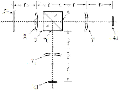
[0047] This embodiment relates to a dual-channel structured light digital phase-contrast microscopic imaging system. The only difference from Embodiment 1 is that the parallel light emitted by the beam expander and collimator unit 2 passes through the ordinary beam splitting prism 8 (other devices can also be used instead ) is reflected onto the object to be measured 5, and its structure schematic diagram is as follows Figure 4 shown.

[0048] The implementation method of the dual-channel structured light digital phase contrast microscopy imaging system described in this embodiment is the same as that in Embodiment 1.

[0049] To sum up, the present invention provides a dual-channel structured light digital phase-contrast microscopic imaging system and its implementation method. The divergent LED light (partially coherent light) is adjusted to parallel light by using a beam expander collimator unit, and then irradiated ( The method is divided into transmission and reflection...


PUM


Abstract

The present invention discloses a dual-channel structure optical digital phase contrast microscopy imaging system and a realization method thereof, wherein the system comprises a light source, a beam expansion collimating unit, a light splitter, a lens group and two same cameras. The beam expansion collimating unit is used to adjust the divergent light emitted out by the light source into the parallel light and irradiate to a to-be-detected object, the light splitter, the lens group and the two cameras form two sets of symmetrical 4f imaging systems and obtain two synchronization images to calculate a real-time bright field micrograph, a differential phase contrast graph and a quantitative phase diagram image. Based on utilizing the common and cheap devices in the market specifically, and by processing simply, a same phase contrast imaging effect can be realized, and the problems that the devices are expensive, and a spatial light modulator is sensitive to the polarization of the light, are solved.

Description

technical field [0001] The invention relates to a dual-channel structured light digital phase-contrast microscopic imaging system and a realization method thereof, belonging to the technical field of microscopic imaging. Background technique [0002] Phase contrast imaging technology is a microscopic imaging technology that can convert the small optical path difference at different positions of the object into intensity difference. Since Zernik invented the phase contrast microscope in 1935 (reference paper: F Zernik. Phase contrast, a new method for the microscopic observation of transparent objects. Physica, Vol.9, No.7), phase contrast microscopy has been widely used At the same time of application, new phase contrast imaging technology is constantly being proposed. The Laura Waller research group of the Berkeley National Laboratory at the University of California, Berkeley proposed a structured light illumination digital phase contrast technology based on array light so...

Claims


Application Information

Patent Timeline
IPC IPC(8): G02B21/36G02B21/14
CPCG02B21/14G02B21/361
Inventor 郑德金王翰林吴郁清李娜娜赵辉顾兆泰安昕张浠
Owner GUANGDONG OPTO MEDIC TECH CO LTD
Why Eureka
Try Eureka