Shear wave elasticity imaging method and device

An elastography and shear wave technology, applied in the field of ultrasonic medical treatment, can solve the problems that the elastic modulus of the blood vessel wall cannot be obtained accurately, and the shear wave elastography cannot be elastic qualitatively and quantitatively, so as to improve the accuracy and applicability of pathological detection. High and wide range of effects

Active Publication Date: 2017-08-18
TSINGHUA UNIV
View PDF6 Cites 24 Cited by
  • Summary
  • Abstract
  • Description
  • Claims
  • Application Information

AI Technical Summary

Problems solved by technology

However, due to some characteristics of some tissues, the shear wave elastography in the related art cannot accurately characterize and quantify the elasticity.
Taking blood vessels as an example, when the distribution of the vessel wall is not circular due to tissue

Method used


Image

  • Shear wave elasticity imaging method and device
  • Shear wave elasticity imaging method and device
  • Shear wave elasticity imaging method and device

Examples


Embodiment Construction

[0024] In order to make the objects, technical solutions and advantages of the present invention more apparent, exemplary embodiments according to the present invention will be described in detail below with reference to the accompanying drawings. Apparently, the described embodiments are only some embodiments of the present invention, rather than all embodiments of the present invention, and it should be understood that the present invention is not limited by the exemplary embodiments described here. Based on the embodiments of the present invention described in the present invention, all other embodiments obtained by those skilled in the art without creative effort shall fall within the protection scope of the present invention.

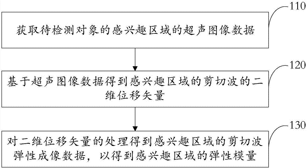
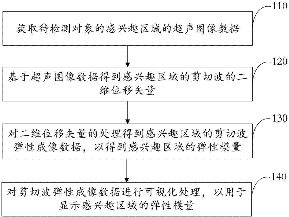
[0025] Acoustic radiation force-based shear wave elastography is an ultrasound elastography technique for assessing tissue stiffness. The basic principle is: the probe emits high-energy ultrasonic waves to the soft tissue of the living body. Under ...


PUM


Abstract

The invention relates to a shear wave elasticity imaging method and device. The method includes the steps: acquiring ultrasonic image data of an interesting area of an object to be detected; acquiring a two-dimensional displacement vector of a shear wave of the interesting area based on the ultrasonic image data; processing the two-dimensional displacement vector to obtain shear wave elasticity imaging data of the interesting area to obtain an elasticity modulus of the interesting area. According to the method, two-dimensional displacement vectors of various moments after excitation of the shear wave are acquired by the aid of a two-dimensional displacement estimation method, the accuracy of an elasticity modulus of the object to be detected is furthest improved, the pathological detection accuracy of the object to be detected is improved, the method is applicable to the object to be detected with a regular shape and also applicable to the object to be detected (such as a blood vessel) with a regular shape caused by influence of peripheral tissues or diseases (such as plaques), elastic quantification can be achieved by the method based on a shear wave, and the method is high in applicability and wider in application range.

Description

technical field [0001] The present invention relates to the field of ultrasonic medical technology, and more particularly to a shear wave elastography method and device. Background technique [0002] Ultrasound imaging is widely used in clinical diagnosis due to its real-time, cheap, non-invasive and non-ionizing radiation advantages. Ultrasound elastography, especially shear wave imaging based on acoustic radiation force, plays a huge role in the qualitative and quantitative measurement of tissue elasticity, for example, vascular shear wave elastography is crucial for the quantification of vessel wall elasticity effect. However, due to some characteristics of some tissues themselves, shear wave elastography in the related art cannot accurately characterize and quantify the elasticity. Taking blood vessels as an example, when the distribution of the vessel wall is not circular due to tissue structure or pathological reasons in the short-axis section of the vessel, the shea...

Claims


Application Information

Patent Timeline
IPC IPC(8): A61B8/00
CPCA61B8/52
Inventor 罗建文何琼曹艳平李国洋
Owner TSINGHUA UNIV
Why Eureka
Try Eureka