
Hybrid multi-mode brain tumour image segmentation method and device

A technology for image mixing and brain tumors, which is applied in the field of medical imaging, can solve the problem of strong dependence on the local optimal initial value of the level set algorithm, and achieve the effects of increasing practicability, speeding up the convergence boundary, and improving accuracy

Inactive Publication Date: 2017-09-12
TIANJIN UNIV
View PDF2 Cites 13 Cited by
  • Summary
  • Abstract
  • Description
  • Claims
  • Application Information

AI Technical Summary

Problems solved by technology

Boundary information can help detect the precise location of the target object, and regional information can prevent boundary leakage, but the level set algorithm cannot solve the problem of being easily trapped in local optimum and strongly dependent on the initial value

Method used


Image

  • Hybrid multi-mode brain tumour image segmentation method and device
  • Hybrid multi-mode brain tumour image segmentation method and device
  • Hybrid multi-mode brain tumour image segmentation method and device

Examples


Embodiment Construction

[0028] 1 Fast FCM theory based on histogram

[0029] The core idea of ​​FFCM is to find the appropriate membership degree and cluster center for the pixel intensity value, so that the variance and iteration error of the cost function within the cluster are minimized. The value of the cost function is the weighted cumulative sum of the 2-norm measure from the pixel to the cluster center. The FFCM clustering and segmentation algorithm is to divide the data into c categories through the fuzzy C-means theory. For an M×N image, suppose {h i ,i=1,2,...,n}, n=M×N, h i is a collection of pixel intensity values ​​in the image histogram. {v j ,j=1,2,…,c} is a set of cluster centers, and μ j (h i ) is h i Belongs to the membership function of class j, so the objective function of FFCM is

[0030]

[0031] and

[0032]

[0033]

[0034] In the formula, ||·|| represents the 2-norm, and b is a constant greater than 1, which controls the ambiguity of the clustering results. to...


PUM


Abstract

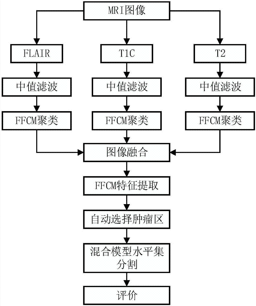
The invention relates to medical apparatuses and medical images and provides an improved hybrid multi-mode brain tumour image segmentation method. A brain tumour area is extracted through employing an FFCM algorithm, a hybrid level set algorithm is utilized to correct a boundary problem existing in the tumour area. The FFCM algorithm and the level set algorithm can be more effectively applied to MRI brain tumour images. The method is characterized by comprising steps that firstly, the MRI images in three modes including T1C, T2 and FLAIR are inputted, median filtering is employed to carry out filtering and initial segmentation of the images to acquire pre-processed images, then linear fusion is employed, lastly, FFCM clustering segmentation is carried out for the fused images, areas with relatively large gray values are automatically extracted, and hybrid level set segmentation is carried out for the acquired tumour under-segmentation areas. The method is mainly applied to medical image acquisition and processing.

Description

technical field [0001] The invention relates to medical equipment, which is an important aspect in the field of medical imaging. It plays an important role in the fields of brain tumor cutting, brain tumor classification, and brain tumor identification. Specifically, it relates to an improved multi-mode brain tumor image hybrid segmentation method and device. Background technique [0002] In recent years, the incidence of brain tumors has been on the rise, accounting for about 5% of systemic tumors and 70% of childhood tumors. In 2015, approximately 23,000 new cases of brain tumors were diagnosed in the United States alone. The uncontrolled and unlimited growth of cells leads to the development of brain tumors. Brain tumors, if not diagnosed and treated early, can cause permanent brain damage and even death. Magnetic Resonance Imaging (MRI) can be used to detect abnormal changes in body tissue and is necessary to determine treatment options for brain tumors. In all treat...

Claims


Application Information

Patent Timeline
IPC IPC(8): G06T7/11G06K9/62
CPCG06T7/11G06T2207/30016G06T2207/30096G06T2207/10088G06F18/23
Inventor 童云飞李锵关欣
Owner TIANJIN UNIV
Learn More