Rapid function magnetic resonance imaging method used for human lung

A technology of functional magnetic resonance and imaging methods, applied in the generation, application, and image enhancement of 2D images, which can solve the problems of increasing the number of phase steps, and achieve the effects of reducing image artifacts, shortening imaging time, and reducing eddy current artifacts

Active Publication Date: 2017-09-12
WUHAN INST OF PHYSICS & MATHEMATICS CHINESE ACADEMY OF SCI
View PDF3 Cites 5 Cited by
  • Summary
  • Abstract
  • Description
  • Claims
  • Application Information

AI Technical Summary

Problems solved by technology

However, the UTE method uses a spatial encoding method in a non-Cartesian co

Method used


Image

  • Rapid function magnetic resonance imaging method used for human lung
  • Rapid function magnetic resonance imaging method used for human lung
  • Rapid function magnetic resonance imaging method used for human lung

Examples


Embodiment Construction

[0046] The specific implementation process and effects of the present invention are given below in conjunction with specific examples.

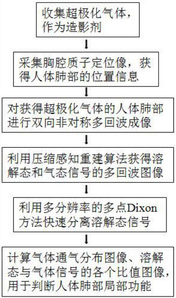
[0047] A fast functional magnetic resonance imaging method for human lungs disclosed by the present invention comprises the following steps:

[0048] Step 1. Collect hyperpolarized gas and use the hyperpolarized gas as a contrast agent. where hyperpolarized gases include 129 Xe or 131 Xe et al. The collected hyperpolarized gas is stored as a gas or as a solid, wherein the solid is sublimated to a gas upon use.

[0049] Step 2. Perform thoracic proton MR positioning imaging on the human lungs to obtain position information of the human lungs. The sequence used for thoracic proton MR localization imaging is FLASH sequence or TSE sequence.

[0050] Step 3. The human body inhales the hyperpolarized gas into the lungs. Inhalation methods include endotracheal intubation, nasal or oral inhalation, etc.

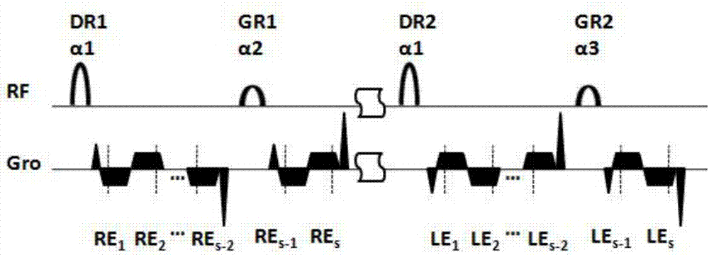
[0051] Step 4, performing two-dimensio...


PUM


Abstract

The invention discloses a rapid function magnetic resonance imaging method used for a human lung. Hyperpolarization gases are collected, and are used as contrast media. Position information of a human lung is acquired. After the hyperpolarization gases are acquired by the human lung, the hyperpolarization gas imaging of the human lung is carried out by adopting a bidirectional asymmetric multi-echo imaging method, and then left and right half-echoes of a plurality of echo times are acquired. The left and right half echoes are combined together to form the full echo data of the corresponding echo time. By adopting a nonlinear conjugate gradient algorithm, the echo images of the dissolved state signals of the various echo times and the echo images of the gaseous state signals of the various echo times are acquired. A B0 field pattern is acquired by using the various echo images of the gaseous signals. Image artifacts caused by the hyperpolarization gases consumed by RF pulses are reduced, and the image artifacts of the dissolved state signals are reduced.

Description

technical field [0001] The invention relates to the technical field of magnetic resonance imaging, in particular to a fast functional magnetic resonance imaging method for human lungs. It is suitable for functional magnetic resonance imaging of human lungs using hyperpolarized gas as a contrast agent, such as functional imaging of lung gas and blood exchange with short sampling time, lung cancer detection, molecular imaging, etc. Background technique [0002] Traditional magnetic resonance imaging (Magnetic Resonance Imaging, MRI) is based on the phenomenon that the observation nucleus in the sample is excited by a radio frequency pulse (RF pulse) in a magnetic field to generate nuclear magnetic resonance. Gradient coils are used to spatially encode the sample. The magnetic resonance signal generated by the sample is received by the electronic system, and the frequency spectrum is transformed to reconstruct the magnetic resonance image. Conventional MRI is mostly used for H...

Claims


Application Information

Patent Timeline
IPC IPC(8): G06T11/00A61B5/055
CPCA61B5/004A61B5/055G06T11/003G06T2207/10088G06T2207/30061G06T2211/412
Inventor 周欣谢军帅李海东张会婷孙献平叶朝辉
Owner WUHAN INST OF PHYSICS & MATHEMATICS CHINESE ACADEMY OF SCI
Why Eureka
Try Eureka