
Tumor image marker extraction method based on gene imaging

An extraction method and marker technology, which can be used in informatics, image enhancement, image analysis, etc., and can solve problems such as undiscovered applications.

Active Publication Date: 2017-09-15
SUZHOU INST OF BIOMEDICAL ENG & TECH CHINESE ACADEMY OF SCI
View PDF4 Cites 10 Cited by
  • Summary
  • Abstract
  • Description
  • Claims
  • Application Information

AI Technical Summary

Problems solved by technology

However, most studies of genetic imaging have focused on the relationship between the two, and its application in the field of imaging markers has not been found

Method used


Image

  • Tumor image marker extraction method based on gene imaging
  • Tumor image marker extraction method based on gene imaging

Examples


Embodiment Construction

[0031] The specific embodiment of the present invention will be described in conjunction with the accompanying drawings.

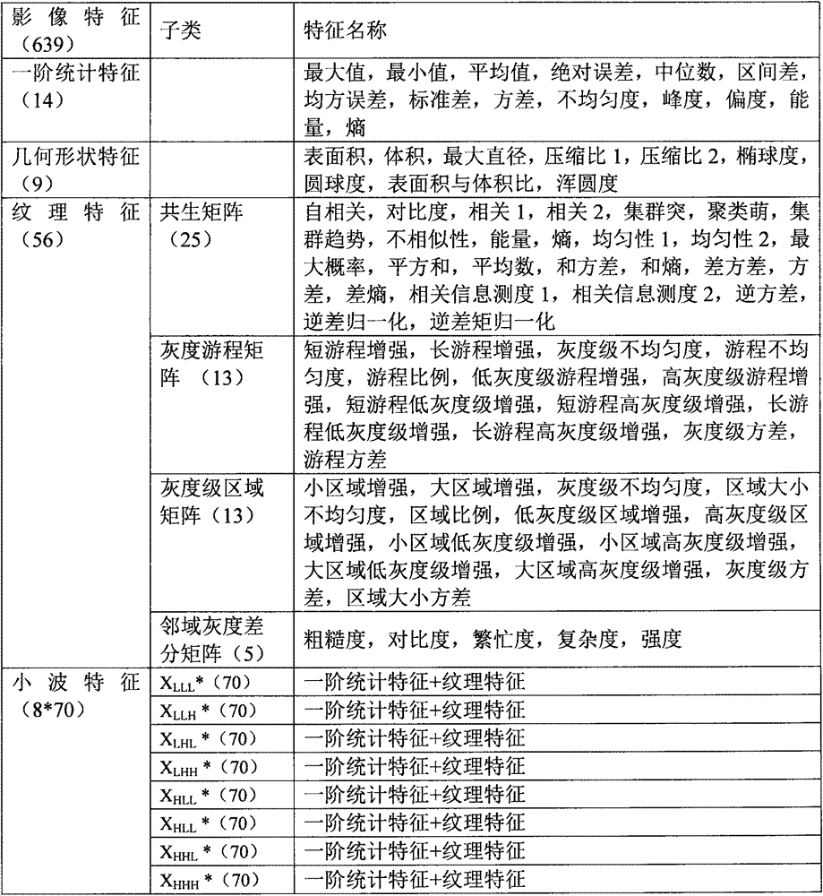
[0032] like figure 1 The flow chart of the method for extracting tumor imaging markers based on genetic imaging is shown. In the method for extracting imaging markers based on genetic imaging in the present invention, first obtain tumor CT data, and then perform tumor CT image analysis to obtain the obtained Spearman correlation was performed between the image features and the prognostic gene modules to obtain the gene image correlation heat map. According to the correlation heat map obtained in the above process and the selected correlation image features, survival analysis and evaluation were performed to obtain a biologically interpretable imaging marker.

[0033] The aforementioned prognostic gene modules are obtained through the following steps. First, the gene expression data is subjected to genome analysis and module clustering. Further, the gene mo...


PUM


Abstract

The invention relates to a tumor image marker extraction method based on gene imaging. The invention provides a non-intrusive biomarker extraction method with high interpretability by adoption of the gene imaging and in combination with the advantages of the imaging and the molecular technology. According to the method, high-dimensional quantitative image features of a tumor CT are extracted and are associated with a corresponding tumor gene expression pattern; and it is assumed that some quantitative image features can reflect a specific gene expression pattern of a tumor and can be used as prognosis markers of the tumor. The ultimate goal is to extract a non-invasive and biologically interpretable imaging marker.

Description

technical field [0001] The invention relates to a method for extracting tumor image markers based on gene imaging, belonging to the field of image markers. Background technique [0002] According to the report of the World Health Organization, cancer is the main cause of global morbidity and death; in my country, malignant tumors have become the number one killer of Chinese residents. The field of tumors is a hot spot in current research, and one of the important challenges is to find tumor markers, which play a vital role in the early diagnosis, prognosis and prognosis of tumors. [0003] Existing tumor marker extraction methods are mainly based on molecular technology. This method requires the use of professional instruments to perform biopsy or surgery on patients, invasively obtain local tumor tissues, and use gene sequencing or protein molecular technology to explore the molecular mechanism of cancer. The root cause of oncogenes, looking for mutation sites and molecula...

Claims


Application Information

Patent Timeline
IPC IPC(8): G06K9/34G06K9/46G06K9/62G06T7/42G06F19/22
CPCG06T7/42G16B30/00G06T2207/30096G06T2207/20064G06V10/267G06V10/443G06V2201/04G06F18/23
Inventor 陈颖高欣
Owner SUZHOU INST OF BIOMEDICAL ENG & TECH CHINESE ACADEMY OF SCI
Learn More