Data processing method and device

A technology of image data and grayscale histogram, applied in the field of medical imaging, can solve the problem of long consumption time

Inactive Publication Date: 2017-10-27
SHANGHAI UNITED IMAGING HEALTHCARE
View PDF5 Cites 21 Cited by
  • Summary
  • Abstract
  • Description
  • Claims
  • Application Information

AI Technical Summary

Problems solved by technology

[0005] In view of this, the embodiment of the present invention provides a data processing method and device to solve the problem of long time consumption in determining the cerebral infarction area and ischemic area in the prior art

Method used


Image

  • Data processing method and device
  • Data processing method and device
  • Data processing method and device

Examples


Embodiment 1

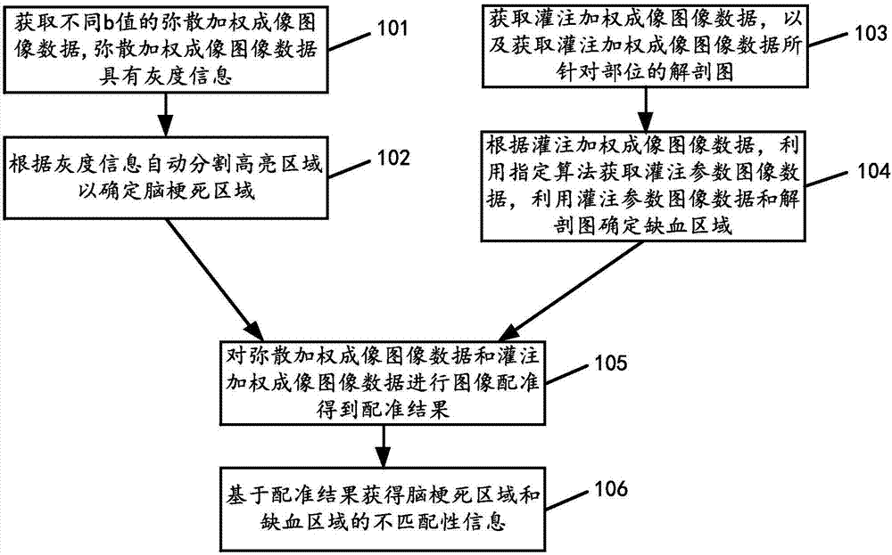
[0068] The embodiment of the present invention provides a data processing method, such as figure 1 As shown, the method includes the following steps:

[0069] 101. Acquire diffusion-weighted imaging image data with different b values, where the diffusion-weighted imaging image data has grayscale information.

[0070] Specifically, DWI image data with different b values ​​can be acquired through MRI imaging technology, where the DWI image data with b value refers to the diffusion-sensitive gradient field parameters applied in the DWI technology.

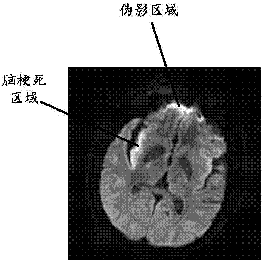
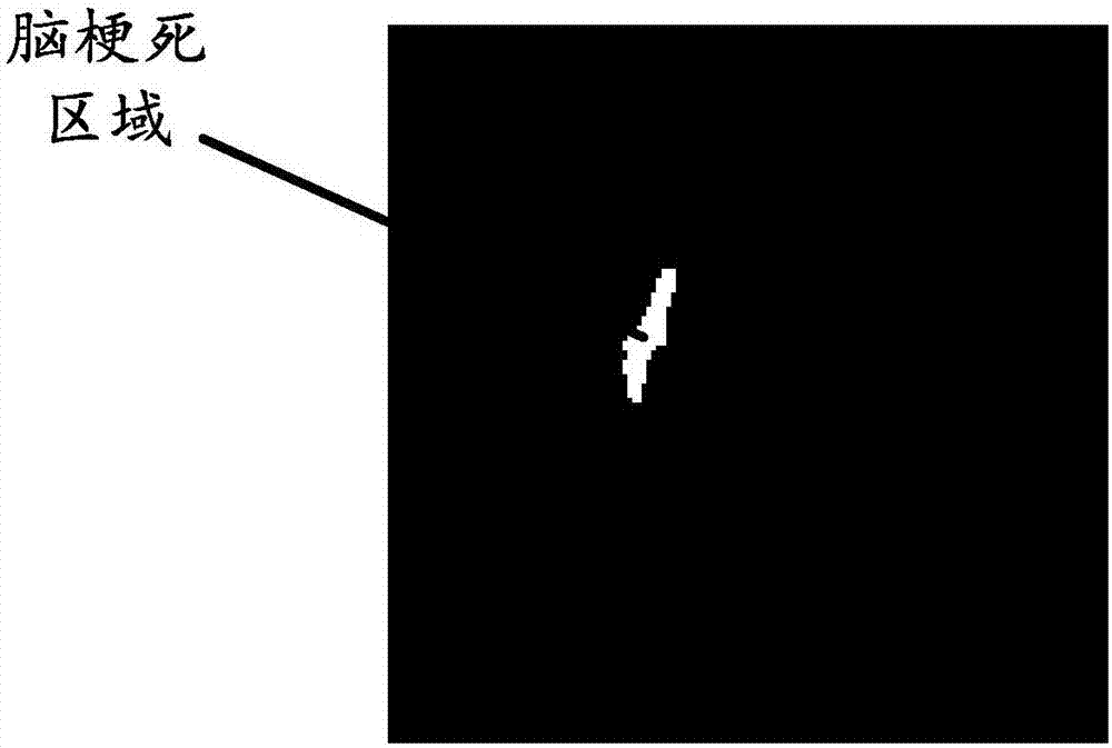
[0071] 102. Automatically segment a highlighted region according to the grayscale information to determine a cerebral infarction region.

[0072] Specifically, since the grayscale information can reflect the grayscale levels corresponding to different regions of the brain, and since the grayscale levels corresponding to cerebral infarction regions are different from those corresponding to normal brain regions, the brain region can be...

Embodiment 2

[0113] An embodiment of the present invention provides a data processing device, such as Figure 8 As shown, the device includes:

[0114] The first acquisition unit 801 is configured to acquire diffusion-weighted imaging image data with different b values, where the diffusion-weighted imaging image data has grayscale information.

[0115] The segmentation unit 802 is configured to automatically segment the highlighted area according to the grayscale information to determine the cerebral infarction area.

[0116] The second acquiring unit 803 is configured to acquire perfusion-weighted imaging image data, and acquire an anatomical map of a site targeted by the perfusion-weighted imaging image data.

[0117] The determining unit 804 is configured to use a specified algorithm to acquire perfusion parameter image data according to the perfusion weighted imaging image data, and determine an ischemic region by using the perfusion parameter image data and the anatomical map.

[01...

Embodiment 3

[0123] The embodiment of the present invention also provides a data processing device, such as Figure 9 As shown, the device includes: a transmitter 91, a receiver 92, a memory 93, and a processor 94 coupled with the memory 93, and the transmitter 91, the receiver 92, the memory 93, and the processor 94 are connected through a bus system Communication; the memory 93 stores a software program that the processor 94 can call to control the transmitter 91 and the receiver 92 . The processor 94 executes the software program for:

[0124] Obtaining diffusion-weighted imaging image data with different b values, the diffusion-weighted imaging image data having grayscale information; automatically segmenting the highlighted region according to the grayscale information to determine the cerebral infarction area; obtaining perfusion-weighted imaging image data, and obtaining the An anatomical map of the part targeted by the perfusion-weighted imaging image data; according to the perfus...


PUM


Abstract

The embodiment of the invention provides a data processing method and device. On one hand, the method comprises the following steps that: according to grey information in obtained diffusion-weighted imaging image data of different b values, automatically segmenting a high-lightness area to determine a cerebral infarction area; according to obtained perfusion weighted imaging image data, utilizing an appointed algorithm to obtain perfusion parameter image data; utilizing the perfusion parameter image data and an obtained anatomy map, determining an ischemic area; carrying out image registration on the diffusion-weighted imaging image data and the perfusion weighted imaging image data; and then, on the basis of a registration result, obtaining the mismatching information of the cerebral infarction area and the ischemic area. In the embodiment of the invention, through the above operations, the cerebral infarction area and the ischemic area can be determined, and since a doctor does not need to analyze the perfusion weighted imaging image data or the weighted imaging image data and does not need to manually determine the cerebral infarction area or the ischemic area, duration consumed for confirming the cerebral infarction area and the ischemic area can be shortened.

Description

【Technical field】 [0001] The invention relates to the technical field of medical imaging, in particular to a data processing method and device. 【Background technique】 [0002] DWI (Diffusion weighted imaging, diffusion weighted imaging) image data and PWI (Perfusion weighted imaging, perfusion weighted imaging) image data can be obtained by using MRI (Magnetic Resonance Imaging, magnetic resonance imaging) imaging technology. The judgment of cerebral infarction provides effective imaging information. In the prior art, after obtaining the DWI image data and the PWI image data, the doctor needs to manually mark the DWI image data and the PWI image data respectively to determine the cerebral infarction area and the ischemic area respectively. [0003] In the course of realizing the present invention, the inventor finds that there are at least the following problems in the prior art: [0004] Since the above operation requires the doctor to manually complete the operation of d...

Claims


Application Information

Patent Timeline
Patent Type & Authority Applications(China)
IPC IPC(8): G06T7/11G06T7/00G06T7/30
CPCG06T7/0012G06T2207/10088G06T2207/30016G06T7/11G06T7/30
Inventor 龙帆马杰延
Owner SHANGHAI UNITED IMAGING HEALTHCARE
Why Eureka
Try Eureka