Method, kit and application of reverse differentiation of blood mononuclear cells to produce human blood-derived autologous retinal stem cells
What is AI technical title?
AI technical title is built by PatSnap AI team. It summarizes the technical point description of the patent document.
A nuclear cell, retinal technology, applied in biomedicine and biological fields, can solve the problems of immune rejection, poor stability, low yield, etc.
Active Publication Date: 2019-03-08
深圳百年干细胞技术研究院有限公司
View PDF4 Cites 0 Cited by
Summary
Abstract
Description
Claims
Application Information
AI Technical Summary
This helps you quickly interpret patents by identifying the three key elements:
Problems solved by technology
Method used
Benefits of technology
Problems solved by technology
In order to solve many problems in the current retinal stem cell preparation method, such as long time-consuming, immune rejection, unsuitable for retinal regeneration therapy, poor stability, high pollution rate, low yield, high risk of carcinogenicity and complicated process
Method used
Image
Click on the blue label to locate the original text in one second.
Reading with bidirectional positioning of images and text.
Examples
Embodiment 1
[0067] Example 1 A set of culture solutions for reverse differentiation of human somatic cells to produce autologous retinal stem cells
[0068] Including cell culture fluid A1, cell culture fluid A2 and cell culture fluid A3.
[0069] It is operated in a safe operating table with a cleanliness level of 10 to 100, and is prepared under low temperature conditions of 4 to 10 ℃.
[0070] Preparation of cell culture fluid A1:
[0071] Add the following ingredients to 500mL DMEM culture medium (purchased from GICO company):
[0072] 10μM RHO kinase inhibitor Y-27632 (purchased from Sigma), 10ng / mL stem cell factor (purchased from R&D company), 10ng / mL interleukin 3 (purchased from R&D company), 10ng / mL interleukin 6 (purchased from R&D company), 10ng / mL interleukin 11 (purchased from R&D company), 10ng / mL macrophage colony stimulating factor (purchased from R&D company), 10ng / mL granulocyte colony stimulating factor (Purchased from R&D Company), 10μg / mL fucoidan, (purchased from Sigma Comp...
Embodiment 2
[0079] Example 2 Kit for preparing autologous retinal stem cells
[0080] It includes the cell culture solution A1, the cell culture solution A2, and the cell culture solution A3 in Example 1, and also includes human somatic cells.
Embodiment 3
[0081] Example 3 Method for preparing retinal stem cells using peripheral venous blood
[0082] 1. Source of blood sample: Aseptically collect peripheral venous blood donated by scientific research staff. Before collecting peripheral venous blood, you should obtain the consent of the blood donor or your immediate family members, and record the genetic and infectious history of the blood donor and your family, as well as all relevant virus examination results in the hospital.
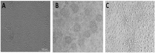
[0083] 2. Separation of mononuclear cells: blood samples are separated using Ficoll standard separation technology to separate mononuclear cells as raw cells. The microscopic view of raw cells is as follows figure 1 -A shown.
[0084] 3. Preparation of retinal stem cells:
[0085] Using the cell culture solution A1, cell culture solution A2, and cell culture solution A3 prepared in Example 1;
[0086] Divide the obtained mononuclear cells into 5x10 6 Density, use cell culture medium A1, at 37°C and 5% CO 2 Cultiv...
PUM
Abstract
The invention belongs to the technical field of biology, in particular relates to the technical field of biomedicine and in particular relates to a cell culture method for enabling human somatic cells to perform reversion differentiation for producing human blood derived auto-retina stem cells as well as a kit and application. The preparation method comprises the following steps: taking somatic cells as raw cells, sequentially culturing the raw cells through a cell culture solution A1, a cell culture solution A2 and a cell culture solution A3, thereby obtaining the blood derived auto-retina stem cells. The human blood cells serve as the raw cells, and the raw cells are subjected to reversion differentiation so as to produce the human blood derived auto-retina stem cells. A cell culture solution formula composed of small molecular substances is used, and the human blood cells are rapidly subjected to reversion differentiation so as to produce the human blood derived auto-retina stem cells under the conditions that the human somatic chromosome DNA sequence is not changed and any foreign gene or DNA fragment is not inserted. The production speed, yield and purity in the invention are obviously superior to those in the prior art.
Description
Technical field [0001] The present invention belongs to the field of biotechnology, in particular to the field of biomedical technology, and more specifically to a cell culture method, kit and application for reverse differentiation of human somatic cells to produce autologous retinal stem cells. Background technique [0002] The retina is a specialized part of the central nervous system located at the fundus of the eye. It originates from the neuroectoderm. It is a light-sensitive and delicate membrane-like structure that forms the basis of various visual functions. The optic cup formed from the neuroectodermal leaf during embryonic development develops. The outer layer of the optic cup forms a single retinal pigment epithelium layer, and the inner layer of the optic cup differentiates into the retinal neurosensory layer. The sensory layers of the retinal nerve from the outside to the inside are: cone, rod layer, outer membrane, outer nuclear layer, outer plexiform layer, inner ...
Claims
Application Information
Patent Timeline
Application Date:The date an application was filed.
Publication Date:The date a patent or application was officially published.
First Publication Date:The earliest publication date of a patent with the same application number.
Issue Date:Publication date of the patent grant document.
PCT Entry Date:The Entry date of PCT National Phase.
Estimated Expiry Date:The statutory expiry date of a patent right according to the Patent Law, and it is the longest term of protection that the patent right can achieve without the termination of the patent right due to other reasons(Term extension factor has been taken into account ).
Invalid Date:Actual expiry date is based on effective date or publication date of legal transaction data of invalid patent.

