
Ultrasound contrast tumor identification method based on multi-mode classifier

A technology of contrast-enhanced ultrasound and classifier, which is applied in the field of image processing, can solve the problems of high recognition accuracy, single classifier, and the inability to achieve high recognition accuracy, and achieve the effect of reducing false positive rate and false negative rate

Inactive Publication Date: 2017-12-22
CHONGQING UNIV
View PDF0 Cites 2 Cited by
  • Summary
  • Abstract
  • Description
  • Claims
  • Application Information

AI Technical Summary

Problems solved by technology

[0003] The other is the use of single-modal data and classification methods, which cannot achieve high recognition accuracy. Although multi-modal data is used in this process, a single classifier is still used for classification, but the multi-modal State data are merged together as input data, which cannot achieve high recognition accuracy

Method used


Image

  • Ultrasound contrast tumor identification method based on multi-mode classifier
  • Ultrasound contrast tumor identification method based on multi-mode classifier
  • Ultrasound contrast tumor identification method based on multi-mode classifier

Examples


Embodiment Construction

[0029] The preferred embodiments of the present invention will be described in detail below with reference to the accompanying drawings.

[0030] An embodiment of the present application provides a multimodal classifier-based method for tumor contrast-enhanced ultrasound identification, the method comprising:

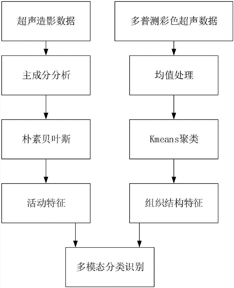
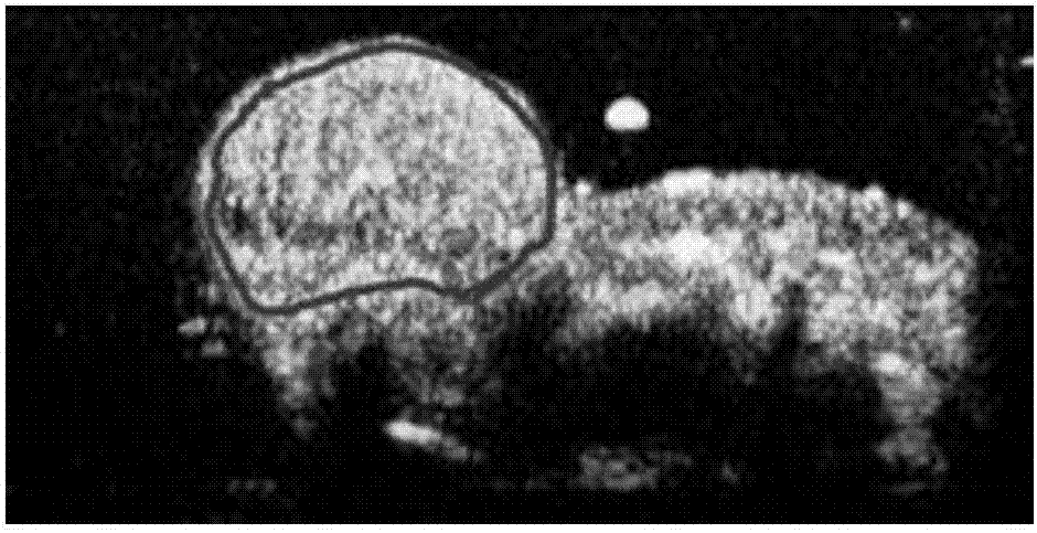
[0031] Pre-acquire ultrasound contrast modal data and Doppler color ultrasound modal data; wherein, the ultrasound modeling modal data is used to characterize the flow of ultrasound contrast agents in blood flow and tissue; the Doppler color ultrasound modal data State data is used to distinguish organizational structures;

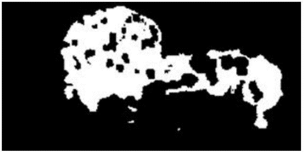
[0032] Preprocessing the contrast-enhanced ultrasound modality data to obtain effective information on the activity of contrast agent microbubbles in blood flow and tissue; preprocessing the Doppler color ultrasound modality data to obtain tissue structure information;

[0033] Perform feature learning on preprocessed CEUS modal data to determine t...


PUM


Abstract

The present invention relates to an ultrasound contrast tumor identification method based on a multi-mode classifier. The method comprises: obtaining ultrasound contrast mode data and Doppler color ultrasound mode data in advance; performing pre-processing of the ultrasound contrast mode data and the Doppler color ultrasound mode data; performing feature learning of the ultrasound contrast mode data through pre-processing to determine the active characteristics of contrast agent microbubbles; performing feature learning of the Doppler color ultrasound mode data through pre-processing to determine different organization structure characteristics; and inputting the active characteristics of the contrast agent microbubbles and the organization structure characteristics into the multi-mode classifier to output the identification result of the tumor organization through the multi-mode classifier. The ultrasound contrast tumor identification method based on the multi-mode classifier can realize detection of the tumor organization and effectively reduce the false negative rate and the false positive rate.

Description

technical field [0001] The invention belongs to the technical field of image processing, and relates to a multimodal classifier-based ultrasound contrast tumor identification method. Background technique [0002] At present, there are few studies on computer-aided diagnosis methods for ultrasound-enhanced tumor diagnosis. Among them, one method is to use B-ultrasound images and images of known liver lesion areas as data, and use human neural networks and decision trees for classification respectively, achieving a certain degree of accuracy. However, due to the diversity of lesion shapes, effective prediction cannot be achieved when dealing with unknown types of lesions. The recognition accuracy rate of this method for liver cancer tissues with different tissue textures is only 65.2%, and the recognition rate for completely different types of liver cancer tissues is only 41.7%. [0003] The other is the use of single-modal data and classification methods, which cannot achie...

Claims


Application Information

Patent Timeline
Patent Type & Authority Applications(China)
IPC IPC(8): G06T7/00G06T7/11G06K9/62
CPCG06T7/0012G06T7/11G06T2207/30096G06T2207/20081G06T2207/10132G06T2207/10024G06F18/23213G06F18/2135G06F18/24155
Inventor 周喜川杨帆赵昕谭跃徐埌唐枋胡盛东林智
Owner CHONGQING UNIV
Learn More