
Endoscope image processing method

An image processing and endoscope technology, applied in the field of endoscope image processing, can solve the problems of too bright area, too dark field of view, uneven lighting of endoscope, etc., and achieve the effect of improving quality

Active Publication Date: 2017-12-26
安徽航天生物科技股份有限公司
View PDF4 Cites 0 Cited by
  • Summary
  • Abstract
  • Description
  • Claims
  • Application Information

AI Technical Summary

Problems solved by technology

[0003] At present, due to the problem of uneven illumination of the endoscope, some areas of the field of view may be too dark and some areas are too bright; When the coupling electronics is in the optimal imaging position, there will be a grid pattern in the image

Method used


Image

  • Endoscope image processing method
  • Endoscope image processing method
  • Endoscope image processing method

Examples


Embodiment Construction

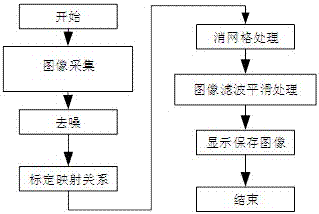
[0031] Such as figure 1 As shown, the present invention provides a kind of endoscopic image processing method, comprises the following steps:

[0032] S1, using Gaussian filtering to remove white noise generated during the image acquisition process;

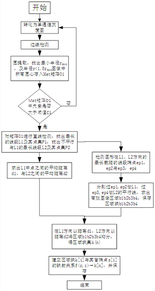
[0033] S2. Calibrate the mapping relationship f(r,c)→k{n} between the original image and the new image, so that the center of the smallest circular bright spot (r, c) in the original image is mapped to the parallelogram area (b1, b2) in the new image , b3, b4), get the block set k{n};

[0034] combine figure 2 As shown, it specifically includes the following sub-steps:

[0035] S2a, converting the denoised RGB image into a grayscale image;

[0036] S2b. Perform adaptive edge extraction on the grayscale image to extract edge information in the image;

[0037] S2c. Perform circle detection on the edge information, find out all circles and the minimum radius r in the edge information min , extract all circles with radius rmin...


PUM


Abstract

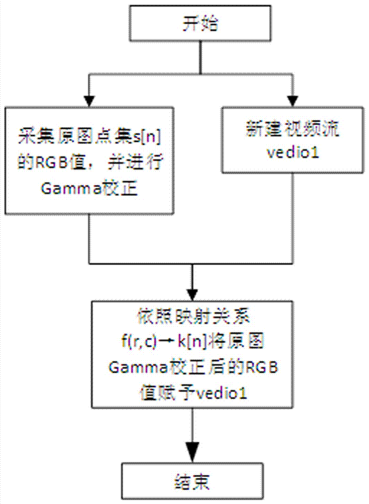
The invention discloses an endoscope image processing method. The method comprises the following steps of S1, using Gaussian filtering to remove a white noise generated during an image acquisition process; S2, calibrating a mapping relation f(r, c)-k{n} of an original image and a newly-constructed image so that a smallest circle bright spot circle center (r, c) in the original image is mapped to a rhomboid area (b1, b2, b3, b4) in the newly-constructed image, and acquiring an area block set k{n}; S3, according to a result of the step2, carrying out grid removing processing on subsequent images; S4, filtering the images acquired from the step3 again so that the images are smooth and continuous; and S5, storing and displaying the processed images. Wavelet transform and a threshold contraction method are used to preprocess collected image information, and through grid removing, gamma correction and filtering smooth processing, image quality is greatly increased.

Description

technical field [0001] The invention relates to the technical field of medical devices, in particular to an endoscope image processing method. Background technique [0002] Endoscope is a detection instrument that integrates traditional optics, ergonomics, precision machinery, modern electronics, mathematics, and software. The endoscope can be used to see lesions that cannot be seen on X-rays, so it is very useful for doctors. [0003] At present, due to the problem of uneven illumination of the endoscope, some areas of the field of view may be too dark and some areas are too bright; When the coupling electronics are in the optimal imaging position, there will be a grid pattern in the image. Contents of the invention [0004] The purpose of the present invention is to provide an endoscope image processing method, which can eliminate the grid in the image displayed by the eyepiece and improve the image quality. [0005] The technical solution adopted by the present inven...

Claims


Application Information

Patent Timeline
Patent Type & Authority Applications(China)
IPC IPC(8): G06T5/00G06T7/13
CPCG06T7/13G06T2207/20024G06T2207/20064G06T2207/10016G06T2207/10024G06T2207/10068G06T5/90G06T5/70
Inventor 李凯王连兵
Owner 安徽航天生物科技股份有限公司
Learn More