Biomarkers for diagnosing vascular stenotic diseases and their uses

A technology for use and vitamins, applied in the field of biomarkers for diagnosing vascular stenosis diseases and its uses, can solve the problem of unreliable speculation of cardiovascular diseases in diabetic patients, and achieve the effect of simple and effective early diagnosis

Active Publication Date: 2020-01-17
BERTIS CO LTD
View PDF1 Cites 0 Cited by
  • Summary
  • Abstract
  • Description
  • Claims
  • Application Information

AI Technical Summary

Problems solved by technology

So far, as the biomarkers, the reduction of adiponectin (adiponectin) secreted from adipocytes, CRP, TNF-α, IL- 6 and other changes in some serum proteins, but the fact is that until now there are no exact biomarkers that can reliably predict the degree of cardiovascular diseases in diabetic patients, especially the degree and disease state of vascular stenosis

Method used


Image

  • Biomarkers for diagnosing vascular stenotic diseases and their uses
  • Biomarkers for diagnosing vascular stenotic diseases and their uses
  • Biomarkers for diagnosing vascular stenotic diseases and their uses

Examples


Embodiment 1

[0066] Example 1: Preparation of patient's blood sample

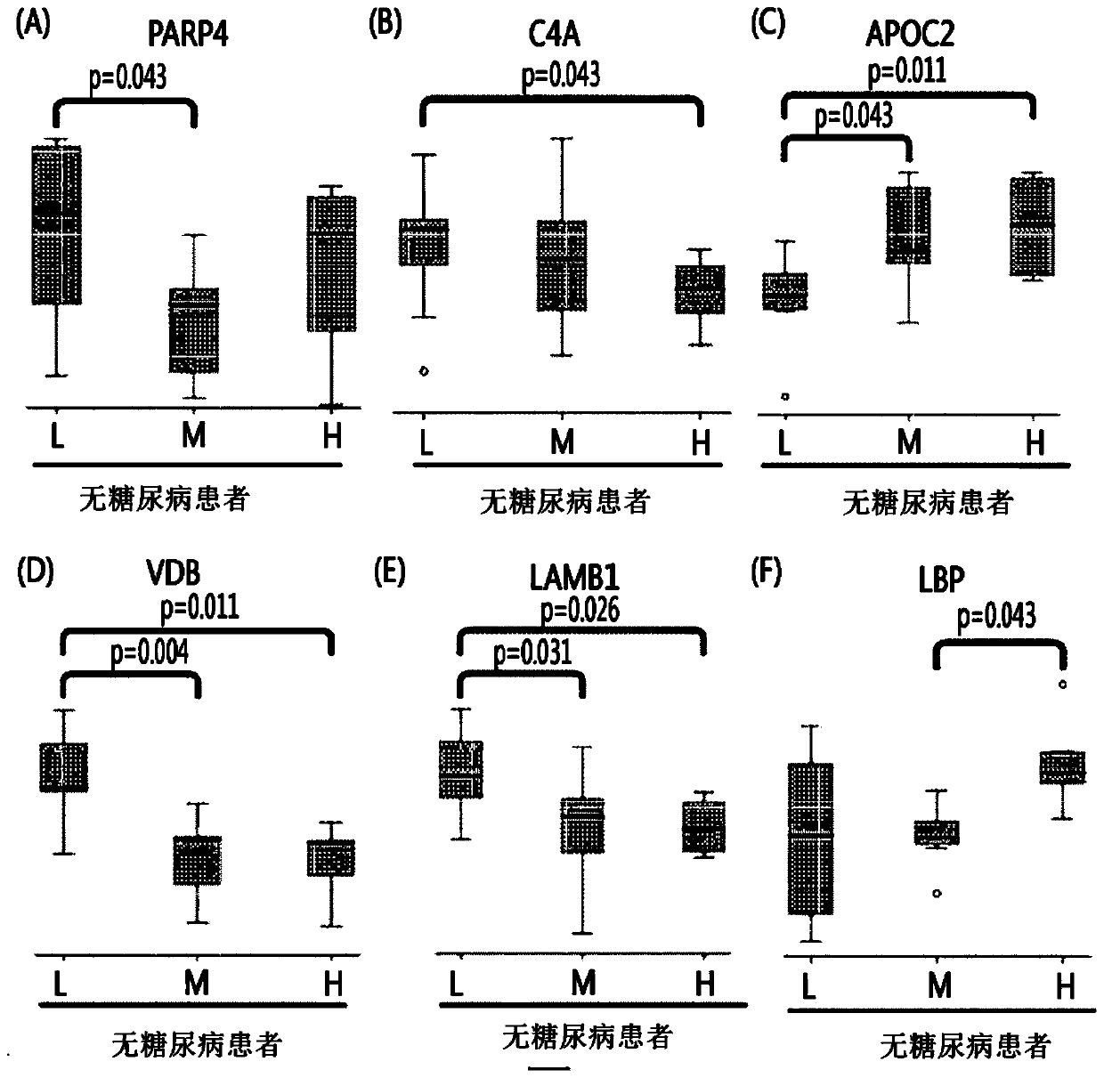
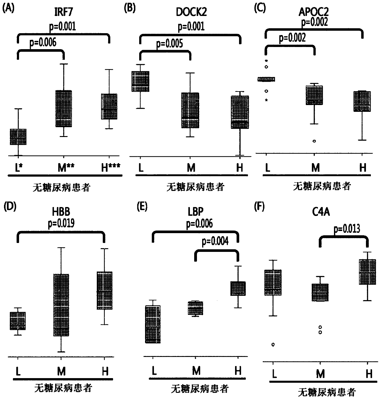
[0067] According to the blockage degree of vascular stenosis, non-diabetic patients were divided into low-risk group patients (0%), medium-risk group patients (0-50%), and high-risk group patients (above 50%). Diabetic patients are divided into low-risk group patients, intermediate-risk group patients, and high-risk group patients. Thereafter, blood samples were collected from these patients.

Embodiment 2

[0068] Example 2: Removal of Abundant Proteins from Patient Blood Samples

[0069] Among the proteins of the blood samples collected in Example 1, 14 high-abundance proteins (albumin, IgG, antitrypsin, IgA, transferrin, haptoglobin, fibrinogen, α2-macrosphere Protein, α1 acid glycoprotein, IgM, apolipoprotein AI, apolipoprotein AII, complement C3, transthyretin) accounted for 95% of the total protein. Therefore, when analyzing the low-abundance proteins of interest, the sensitivity of these 14 high-abundance proteins is greatly reduced, so the MARS (Multiple Affinity Removal System (Multiple Affinity Removal System)) column (P / N 5188 -6558, Agilent) to remove the 14 proteins, and only the remaining proteins were used for subsequent analysis.

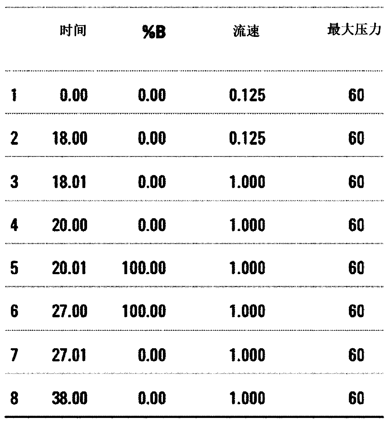
[0070] Specifically, the process of removing high-abundance proteins using MARS (Multiple Affinity Removal System) column (P / N 5188-6558, Agilent) is as follows.

[0071] 2 μl of 50x protease inhibitor (Roche) was added to 40 μl of the...

Embodiment 3

[0077] Example 3: Peptidation of protein samples

[0078] In order to perform analysis with a mass spectrometer, a process of cleavage of protein in peptide units by an enzyme (trypsin) is required first, so peptide cleavage of protein is carried out by the following process.

[0079] 150 μl of DTT solution (final concentration: 6M urea, 10 mM DTT, 50 mM tris) was added to 50 μl of blood sample with a concentration of 60 μg to remove abundant protein, and reacted (reduced) at 37° C. for 1 hour. After treating 20 μl of IAA solution (final concentration: 5.45 M of urea, 50 M of IAA, 50 mM tris) in 200 μl of the reacted (reduced) sample, reacted (alkylated) at room temperature for 30 minutes in the dark, Then, 1 ml of Tris solution (50 mM, pH 8) (final concentration: 1 M urea, 50 mM tris, final volume: 1.2 ml) was added to the sample. Thereafter, the trypsin solution was dispensed in units of 5 μl and reacted at 37° C. for 12 hours.


PUM


Abstract

Provided are: a composition for diagnosing non-diabetic angiostenosis, containing a preparation measuring the level of one or more proteins selected from the group consisting of poly (ADP-ribose) polymerase 4 (PARP4), vitamin D-binding protein (VDB), and laminin subunit beta-1 (LAMB1); a composition for diagnosing diabetic angiostenosis, containing a preparation measuring the level of one or more proteins selected from the group consisting of interferon regulatory factor 7 (IRF7), a dedicator of cytokinesis protein 2 (DOCK2), and a hemoglobin subunit beta (HBB); a kit for diagnosing angiostenosis, containing the composition for diagnosing diabetic angiostenosis; a method for providing information for diagnosing non-diabetic angiostenosis; and a method for providing information for diagnosing non-diabetic angiostenosis. The present invention provides a composition for diagnosing angiostenosis, so as to measure and compare the changing protein expression levels in non-diabetic or diabetic angiostenosis patients, thereby enabling early diagnosis of angiostenosis and a meaningful prediction or an identification of the degree of the disease. In addition, the diagnostic composition of the present invention enables non-invasive diagnosis, thereby enabling simple and valid early stage diagnosis of angiostenosis by using blood and urine testing and the like.

Description

technical field [0001] The present invention provides a composition for diagnosing non-diabetic vascular stenosis, a composition for diagnosing diabetic vascular stenosis, a kit for diagnosing vascular stenosis comprising the composition, and a diagnostic kit for diagnosing vascular stenosis. A method of providing information on non-diabetic vascular stenotic disease and a method of providing information for diagnosing diabetic vascular stenotic disease. Background technique [0002] Recently, there has been a rapid increase in the number of patients with metabolic syndrome who manifest various diseases complexly due to urbanization of the living environment, excessive nutritional intake, and the like. Globally, the risk of diabetes and obesity as metabolic diseases increases, and the prevalence of diabetes has begun to exceed 10% of the population. [0003] According to the results of the Korean National Health and Nutrition Survey from 2005 to 2007, 10% of Korean adults a...

Claims


Application Information

Patent Timeline
Patent Type & Authority Patents(China)
IPC IPC(8): C07K16/00G01N33/68
CPCC12Q1/68G01N33/68
Inventor 金光杓崔诚希李厚槿李祥原
Owner BERTIS CO LTD
Why Eureka
Try Eureka