CNN-based multimodal nasopharyngeal tumor joint segmentation method

A combined segmentation and nasopharyngeal technology, applied in image analysis, biological neural network model, image data processing, etc., can solve the problem of difficult to meet high-precision clinical needs, unsatisfactory research results, and little research on image segmentation, etc. problem, to achieve the effect of shortening the segmentation time, simple structure, and improving the segmentation accuracy

Active Publication Date: 2018-01-19
SICHUAN UNIV
View PDF7 Cites 9 Cited by
  • Summary
  • Abstract
  • Description
  • Claims
  • Application Information

AI Technical Summary

Problems solved by technology

However, it turns out that single-modal CT and RT images contain very limited information. In fact, nasopharyngeal tumors are difficult to have obvious boundaries even in enhanced CT images, so only single-modal CT and MR images are used. Difficult to meet high-precision clinical needs
[0005] Nasopharyngeal carcinoma is one of the high-incidence tumor

Method used


Image

  • CNN-based multimodal nasopharyngeal tumor joint segmentation method
  • CNN-based multimodal nasopharyngeal tumor joint segmentation method
  • CNN-based multimodal nasopharyngeal tumor joint segmentation method

Examples


Embodiment Construction

[0033] A detailed description will be given below in conjunction with the accompanying drawings.

[0034] In order to make the object, technical solution and advantages of the present invention clearer, the present invention will be further described in detail below in combination with specific embodiments and with reference to the accompanying drawings. It should be understood that these descriptions are exemplary only, and are not intended to limit the scope of the present invention. Also, in the following description, descriptions of well-known structures and techniques are omitted to avoid unnecessarily obscuring the concept of the present invention.

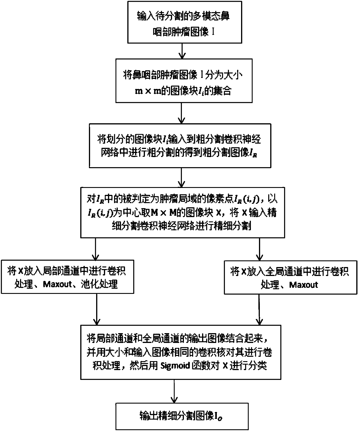
[0035] figure 1 It is an algorithm flow chart of the segmentation method of the present invention. Combine now figure 1 A CNN-based multimodal nasopharyngeal tumor joint segmentation method proposed by the present invention is described in detail. The segmentation method of the present invention includes:

[0036] Step 1...


PUM


Abstract

The present invention relates to a CNN-based multimodal nasopharyngeal tumor joint segmentation method. The method comprises: segmenting a multimodal nasopharyngeal tumor image into image blocks of the same size; inputting the segmented image blocks into a rough segmentation CNN for rough segmentation to obtain rough segmented images, and obtaining an image block determined as a tumor area by rough segmentation processing; inputting the rough segmented images into the fine CNN for fine segmentation; and carrying out fine segmentation on each pixel in the image blocks determined as a tumor area, determining the category of the pixel, and obtaining pixel-level finely segmented images finally. The segmentation method of the present invention does not need to determine the category of all thepixels in the nasopharyngeal tumor image, and only needs to determine a few lesion areas, thereby greatly improving the efficiency of the segmentation algorithm and shortening the segmentation time;and in addition, both multimodal data and CNN are used for image segmentation, and richer information of the to-be-segmented tumor image is preserved, so that the segmentation accuracy of the algorithm is improved.

Description

technical field [0001] The invention belongs to the field of tumor image segmentation, and in particular relates to a CNN-based multimodal nasopharyngeal tumor joint segmentation method. Background technique [0002] In the field of medical image segmentation, the current research results on head and neck tumor image segmentation algorithms are increasingly fruitful. Especially in the image segmentation method of head tumors, good segmentation results have been achieved. Nasopharyngeal tumor is a common head and neck tumor. According to the statistics of the World Health Organization, about 80% of nasopharyngeal cancers in the world occur in my country. Therefore, it is very useful to find an effective image segmentation method for nasopharyngeal tumors. meaningful. At present, there are few research results on nasopharyngeal carcinoma at home and abroad. The existing nasopharyngeal tumor image segmentation algorithms are either too complicated in method and difficult to be...

Claims


Application Information

Patent Timeline
IPC IPC(8): G06T7/11G06N3/04
Inventor 王艳胡光亮何嘉吴锡周激流
Owner SICHUAN UNIV
Why Eureka
Try Eureka