Schematic eye for fundus detection and using method thereof

A model eye and film technology, applied in the field of model eye, can solve the problem of not considering the state of the human eye, and achieve the effect of simple structure and good versatility

Pending Publication Date: 2018-02-02
浙江省医疗器械检验院
View PDF0 Cites 7 Cited by
  • Summary
  • Abstract
  • Description
  • Claims
  • Application Information

AI Technical Summary

Problems solved by technology

This method is measured in the air and does not take into accou

Method used


Image

  • Schematic eye for fundus detection and using method thereof
  • Schematic eye for fundus detection and using method thereof
  • Schematic eye for fundus detection and using method thereof

Examples


Embodiment Construction

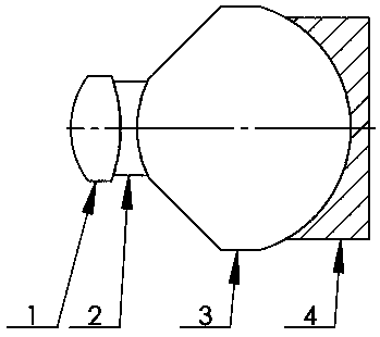
[0021] The application will be further described below in conjunction with the accompanying drawings. figure 1 Middle left is front, right is back.

[0022] refer to figure 1 It is a model eye for fundus testing. The model eye is composed of three optical lenses, a film 4 with a field angle scale and a resolution line pair. The entire lens is sequentially the first lens 1, the second lens 2, the third lens 3, and the negative film 4 from the object side to the image side. The adjacent surface is close to each other, and the field angle scale and the resolution line pair are arranged on the adjacent surface of the negative film 4 and the third lens 3 (the arc surface at the front of the negative film 4 ).

[0023] The first lens 1 is a convex lens, the radii of the front surface and the back surface are 8 and -14.027 respectively, the center thickness is 4.5, and the material is K9 glass; the second lens 2 is a concave lens, and the radii of the front surface and the back su...


PUM


Abstract

The invention designs a model eye used for fundus detection and a using method thereof. The model eye is composed of three optical lenses, a film with a field angle scale and a resolution line pair. The model eye is the first lens, the second lens, the third lens, and the film in order from the object side to the image side. Adjacent surfaces of the first lens, the second lens, the third lens and the film are in close contact with each other, and the field angle scale and the resolution line pair are arranged on the arc surface of the front part of the film. The invention also includes a method of using the model eye. The optical structure and the refractive index difference between optical lenses or lens groups in the present invention simulate the actual refractive system of the human eye, and the field of view scale and resolution line pair carved on the film are based on the index requirements in ISO10940, and replace those in ISO10940 The method is used to detect the field of view and resolution of fundus imaging inspection medical devices represented by fundus cameras.

Description

technical field [0001] The present application relates to a model eye for fundus detection, which is suitable for use as a model eye to detect the performance indicators of fundus imaging inspection medical devices represented by fundus cameras. Background technique [0002] Fundus imaging inspection medical devices mainly include fundus cameras, fundus imaging machines, fundus scanners, etc. Among them, the fundus camera is a typical representative, which records and saves the fundus in the form of black and white or color photos, so as to diagnose fundus diseases. Its main optical properties include resolution, field of view, etc. The accuracy of these performance indicators is directly related to the effectiveness of diagnosis, and further to the safety of patients' eyes. The international standard IS010940 "Fundus Camera for Ophthalmic Instruments" for fundus camera products has been released. Currently, the resolution, field of view and other measurement methods in the...

Claims


Application Information

Patent Timeline
IPC IPC(8): A61B3/12A61B3/14A61B3/00G09B23/28
CPCA61B3/0025A61B3/12A61B3/14G09B23/28
Inventor 贾晓航彭建华王敬涛
Owner 浙江省医疗器械检验院
Why Eureka
Try Eureka