

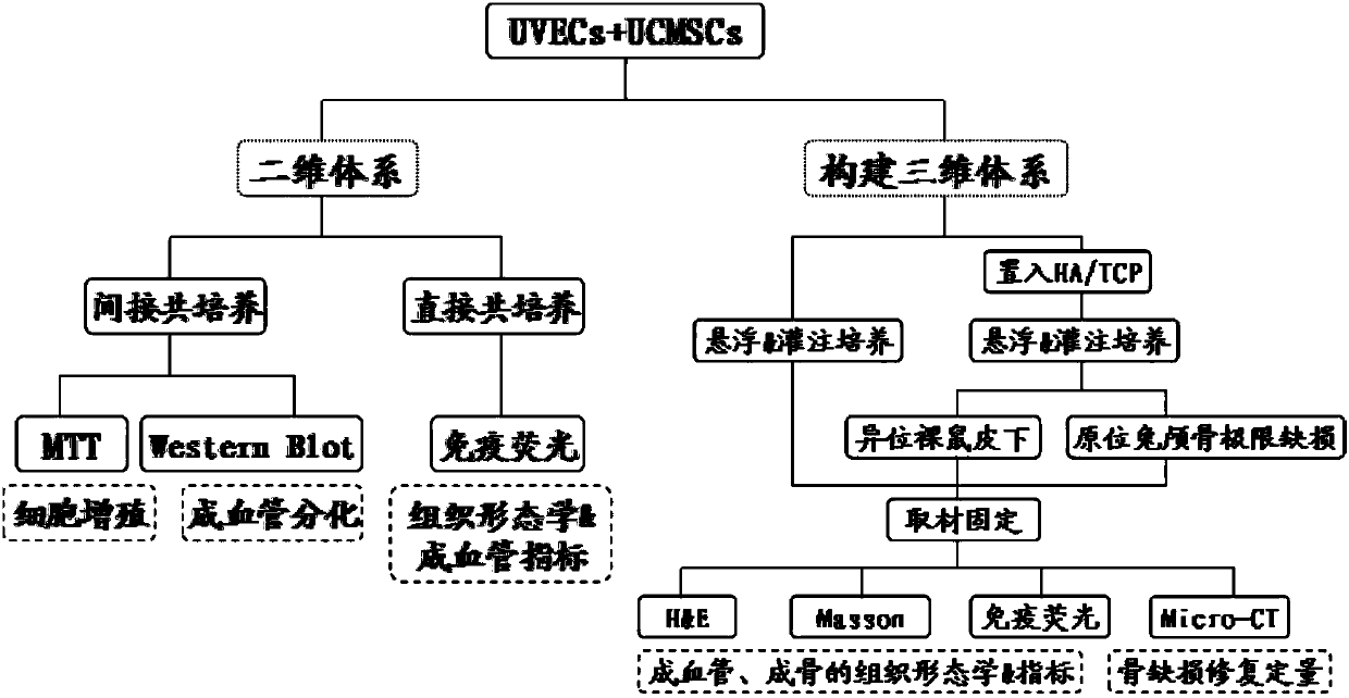
Experimental method for promoting tissue engineering pre-vascularization by umbilical cord mesenchymal stem cells
A technology of stem cells and tissue engineering, applied in the medical field, can solve problems such as unclear
- Summary
- Abstract
- Description
- Claims
- Application Information
AI Technical Summary
Problems solved by technology
Method used
Image


Examples
Embodiment
[0142] 1. Primary cell culture of umbilical cord mesenchymal stem cells. Such as figure 2 shown.
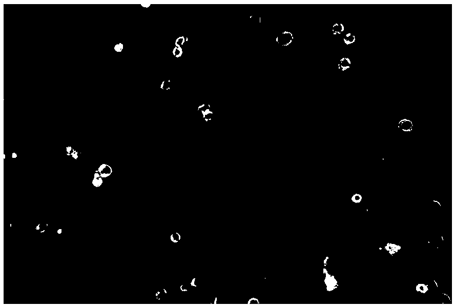
[0143] 2. Primary culture of umbilical cord vein endothelial cells. Such as image 3 shown.
[0144] 3. Detection of surface markers of umbilical cord mesenchymal stem cells. Such as Figure 4-Figure 10 shown.
[0145] 4. Detection of osteogenic differentiation of umbilical cord mesenchymal stem cells. Such as Figure 11 shown.
[0146] 5. Detection of adipogenic differentiation of umbilical cord mesenchymal stem cells. Such as Figure 12 shown.
[0147] 6. Staining of the colony formation rate of umbilical cord mesenchymal stem cells. Such as Figure 13 shown.
[0148] 7. The umbilical cord vein endothelial cells positively expressed CD-31. Such as Figure 14 shown.
[0149] 8. The umbilical cord vein endothelial cells positively expressed VEGF. Such as Figure 15 shown.
[0150] 9. Umbilical cord vein endothelial cells positively expressed factor Ⅷ. Such as ...
PUM
 Abstract
Description
Claims
Application Information