Eureka AIR delivers breakthrough ideas for toughest innovation challenges, trusted by R&D personnel around the world.

Hysteroscope

A technology of hysteroscope and insertion tube, applied in the field of hysteroscope, can solve the problems of complicated operation, unreasonable setting, and complicated use of hysteroscope, and achieve the effects of low production cost, good practical value and simple structure

Pending Publication Date: 2018-03-20
SHANGHAI ANQING MEDICAL INSTR
View PDF0 Cites 8 Cited by
  • Summary
  • Abstract
  • Description
  • Claims
  • Application Information

AI Technical Summary

Problems solved by technology

[0010] The operation of this kind of hysteroscope is relatively complicated in actual use, and its safety and reliability are low.
[0011] In the existing technology taking the above-mentioned hysteroscope as an example, most hysteroscopes have the disadvantages of many operating parts and poor sealing performance, and the setting of various parts on the handle of the hysteroscope is not very reasonable. Inconvenient in the process
[0012] In addition, due to the high production cost of hysteroscopes in the prior art, it needs to be used repeatedly. In addition to the need for disinfection before each use, the use is relatively cumbersome, and the probability of cross-infection between patients is also very high.

Method used


Image

  • Hysteroscope
  • Hysteroscope
  • Hysteroscope

Examples


no. 1 example

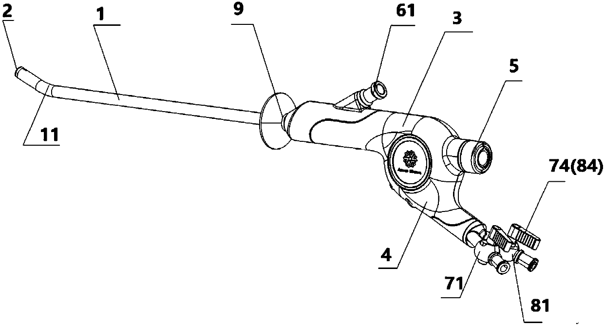
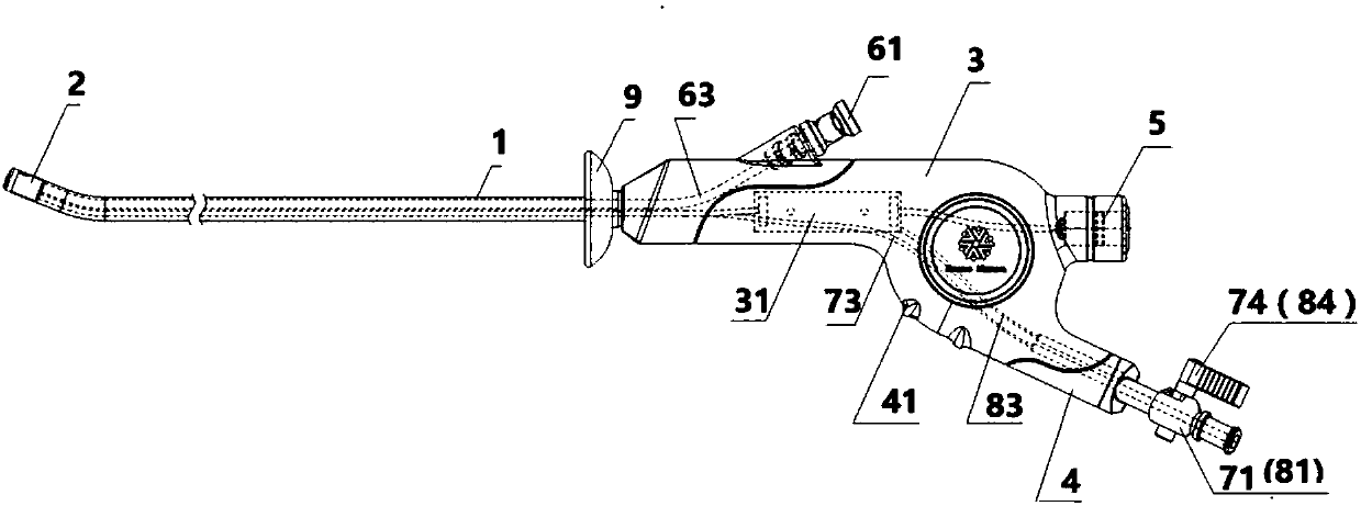
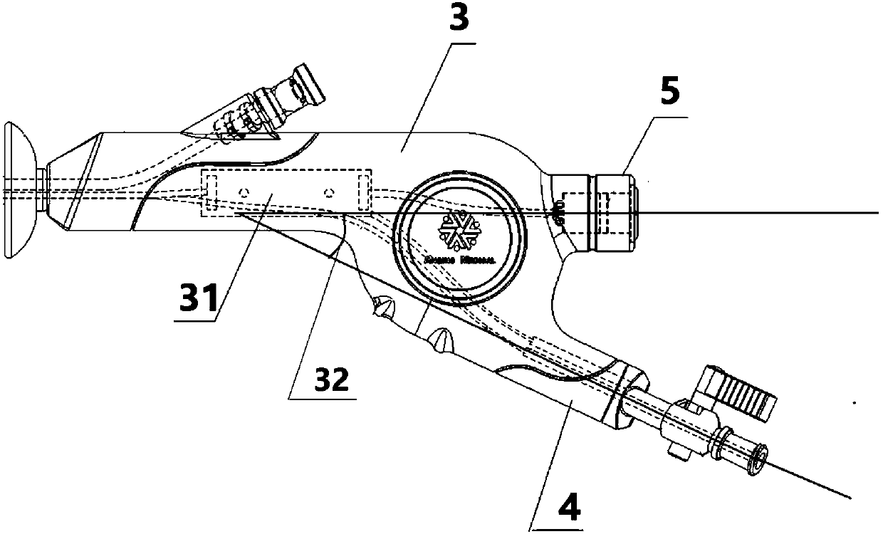
[0045] Such as figure 1 and Figure 5-1As shown in , the first embodiment of the present invention discloses a hysteroscope, including: an insertion part, the insertion part includes an insertion tube 1 and an imaging module seat 2 fixed to the front end of the insertion tube, and the imaging module seat 2 is provided with The optical imaging device 21 and the light source 22; the operating part, the operating part is connected to the end of the insertion part through a rigid connector, including a handle 3 and a handle 4 at a certain angle with the handle, the handle 3 and the handle 4 together form a pistol shape, the handle 3 is provided with a self-locking quick connector 5 electrically connected to the optical imaging device 21 and the light source 22; wherein, as figure 1 , diagram 2-1 and Figure 5-1 As shown in , the handle is provided with an instrument mouth 61, and the front end surface of the insertion part is provided with an instrument outlet 62. The instrume...

no. 2 example

[0066] Such as figure 1 As shown in , the second embodiment of the present invention discloses a hysteroscope, including: an insertion part, the insertion part includes an insertion tube 1 and an imaging module seat 2 fixed to the front end of the insertion tube, and the imaging module seat 2 is provided with The optical imaging device 21 and the light source 22; the operating part, the operating part is connected to the end of the insertion part through a rigid connector, including a handle 3 and a handle 4 at a certain angle with the handle, the handle 3 and the handle 4 together form a pistol shape, the handle 3 is provided with a self-locking quick connector 5 electrically connected to the optical imaging device 21 and the light source 22; wherein, as figure 1 , Figure 2 and Figure 5-2 As shown in , the handle is provided with an instrument mouth 61, and the front end surface of the insertion part is provided with an instrument outlet 62. The instrument mouth 61 communic...

no. 3 example

[0081] Such as image 3 and Figure 5-1As shown in , the third embodiment of the present invention discloses a hysteroscope, including: an insertion part, the insertion part includes an insertion tube 1 and an imaging module seat 2 fixed to the front end of the insertion tube, and the imaging module seat 2 is provided with The optical imaging device 21 and the light source 22; the operating part, the operating part is connected to the end of the insertion part through a rigid connector, including a handle 3 and a handle 4 at a certain angle with the handle, the handle 3 and the handle 4 together form a pistol shape, the handle 3 is provided with a self-locking quick connector 5 electrically connected to the optical imaging device 21 and the light source 22; wherein, as image 3 and Figure 5-1 , 5-2 As shown in , the handle is provided with an instrument mouth 61, and the front end surface of the insertion part is provided with an instrument outlet 62. The instrument mouth ...


PUM

PropertyMeasurementUnit
Diameteraaaaaaaaaa

Abstract

The invention discloses a hysteroscope, which includes an insert portion comprising an insert tube and an imaging module seat which is provided with an optical images acquisition device and a light source; the hysteroscope also includes an operation department, which is connected with the end of the insert portion and includes a handle and a handgrip with an angle to the handle. The handle is provided with an instrument entry, the front end of the insert portion is provided with an instrument export and the instrument entry is connected with the instrument export through an instrument channelinserted inside the handle and inserted into the inner cavity of the insert tube; the handgrip is provided with an inflow base and an outflow base, the front end of the insert portion is provide witha water intake and an outlet, and the inflow base and the outflow base are separately connected with the water intake and the outlet through an inflow channel and an outflow channel inserted inside the handle and inserted into the inner cavity of the insert tube. The hysteroscope can be used in the routine examination of the patients' uterus, and is convenient and safe to treat patients in time; at the same time, the whole process of examination and treatment is visible, safe and reliable, minimizes the damage to the patients, and has a high practical value.

Description

technical field [0001] The invention relates to the field of medical instruments, in particular to a hysteroscope. Background technique [0002] Hysteroscope is a kind of light source hysteroscope mainly used for examination and treatment of uterine cavity. It uses the front part of the mirror body to enter the uterine cavity. It has become the preferred inspection method for gynecological hemorrhagic diseases and intrauterine lesions with intuitiveness and accuracy. However, in the prior art, when examining the patient's uterus, it is usually necessary to extend the hysteroscope from the patient's body, enter the uterus, and observe the patient. treatment, or keep the hysteroscope in the patient's body, insert the treatment instrument into the patient's body at the same time, and treat the patient. [0003] Existing Embodiment 1 discloses a hysteroscope, including a mirror sheath and a speculum, the speculum is arranged in the mirror sheath, an inner sheath and an outer sh...

Claims


Application Information

Patent Timeline
IPC IPC(8): A61B1/303A61B1/015A61B1/018A61B1/04A61B1/06
CPCA61B1/00066A61B1/015A61B1/018A61B1/042A61B1/0684A61B1/303
Inventor 不公告发明人
Owner SHANGHAI ANQING MEDICAL INSTR
Why Eureka
Eureka Blog
Learn More