Image classification method, device, storage medium and equipment

A classification method and image technology, applied in the field of machine learning, can solve problems such as low accuracy of machine learning models, ignoring lung nodules, and affecting the accuracy of lung cancer attribute recognition, achieving high accuracy, high classification accuracy, The effect of accurate detection

Active Publication Date: 2018-04-10
TENCENT TECH (SHENZHEN) CO LTD +1
View PDF4 Cites 15 Cited by
  • Summary
  • Abstract
  • Description
  • Claims
  • Application Information

AI Technical Summary

Problems solved by technology

[0005] In the CT image of the lung lesion area, the size distribution of pulmonary nodules is wide. Using single-scale features for model training will lead to low accuracy of the trai...

Method used


Image

  • Image classification method, device, storage medium and equipment
  • Image classification method, device, storage medium and equipment
  • Image classification method, device, storage medium and equipment

Examples


Embodiment Construction

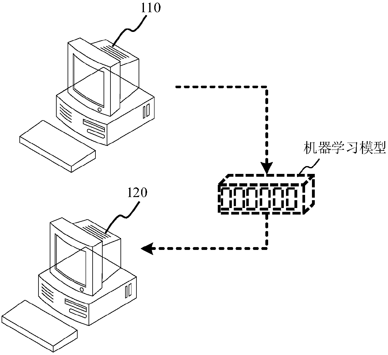
[0033] In order to make the object, technical solution and advantages of the present invention clearer, the implementation manner of the present invention will be further described in detail below in conjunction with the accompanying drawings.

[0034] Before explaining and describing the embodiments of the present invention in detail, some terms involved in the embodiments of the present invention will be explained first.

[0035] 3D Imaging: Refers to medical imaging images of diseased organs. Wherein, the three-dimensional imaging map can be aimed at the whole diseased organ, or only at the lesion area of ​​the diseased organ.

[0036] In addition, the three-dimensional imaging image may be either a CT image or an MRI image, which is not specifically limited in this embodiment of the present invention. For example, a 3D imaging image may be composed of multiple 2D CT images of different slice layers.

[0037] Sensitivity: Among all the medical imaging images whose disease...


PUM


Abstract

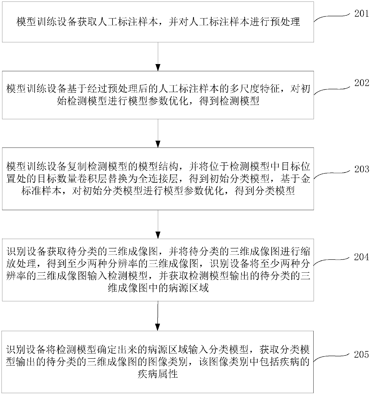
The invention discloses an image classification method, a device, a storage medium and equipment, and belongs to the technical field of machine learning. The method comprises the following steps of acquiring a to-be-classified three-dimensional imaging graph, performing scaling treatment on the to-be-classified three-dimensional imaging graph, and inputting obtained three-dimensional imaging graphs of at least two resolution ratios into a detection model, wherein the detection model is obtained through the machine learning process based on the multi-scale features of a manual labeling sample;acquiring a disease source region in the to-be-classified three-dimensional imaging graph output by the detection model; inputting the determined disease source region into a classification model, wherein the classification model is obtained through the machine learning process on the basis of a gold standard sample, and the gold standard sample is an image sample which is used for correctly distinguishing the attributes of disease sources; acquiring the graph category of the to-be-classified three-dimensional imaging graph outputted by the classification model, wherein the graph category comprises the disease attributes of diseases. According to the invention, the model is high in accuracy after being trained based on the multi-scale features and the gold standard sample. Meanwhile, the accuracy of graph classification is greatly improved.

Description

technical field [0001] The present invention relates to the technical field of machine learning, in particular to an image classification method, device, storage medium and equipment. Background technique [0002] As the core of artificial intelligence, machine learning technology has been applied in various fields, such as the medical field. In the medical field, the use of machine learning techniques to classify medical imaging images can usually identify disease attributes. Among them, the disease attribute can generally be divided into benign (non-disease) and malignant (disease). Taking lung cancer as an example, machine learning technology is currently used to classify medical imaging images of lung lesions to identify whether lung cancer is benign or malignant. [0003] Continuing to take lung cancer as an example, when the relevant technology uses image classification to identify benign and malignant lung cancer, it first uses a pre-trained machine learning model t...

Claims


Application Information

Patent Timeline
IPC IPC(8): G06T7/00G06T3/40G06T17/00G06N3/04G06N3/08
CPCG06N3/08G06T3/40G06T7/0012G06T17/00G06T2207/10081G06T2207/30064G06N3/045
Inventor 孙星曹鸿吉郭晓威
Owner TENCENT TECH (SHENZHEN) CO LTD
Why Eureka
Try Eureka