Head-mounted micro light sheet microscope

A microscope and head-mounted technology, applied in the field of fluorescence imaging, can solve problems such as unfavorable brain neural activity, limit animal activity range, research, etc., achieve good optical layer selection ability, reduce phototoxicity, improve imaging speed and signal-to-noise. the effect of

Inactive Publication Date: 2018-04-27
SOUTHERN MEDICAL UNIVERSITY
View PDF6 Cites 6 Cited by
  • Summary
  • Abstract
  • Description
  • Claims
  • Application Information

AI Technical Summary

Problems solved by technology

Many studies have shown that many neural activities in the animal brain will be affected during anesthesia, which is not conducive to the study of brain neural activities in a normal state. Some imaging devices limit the range of activities of animals, making it impossible to perform certain experiments or measurements on animals. certain parameters

Method used


Image

  • Head-mounted micro light sheet microscope
  • Head-mounted micro light sheet microscope

Examples


Embodiment 1

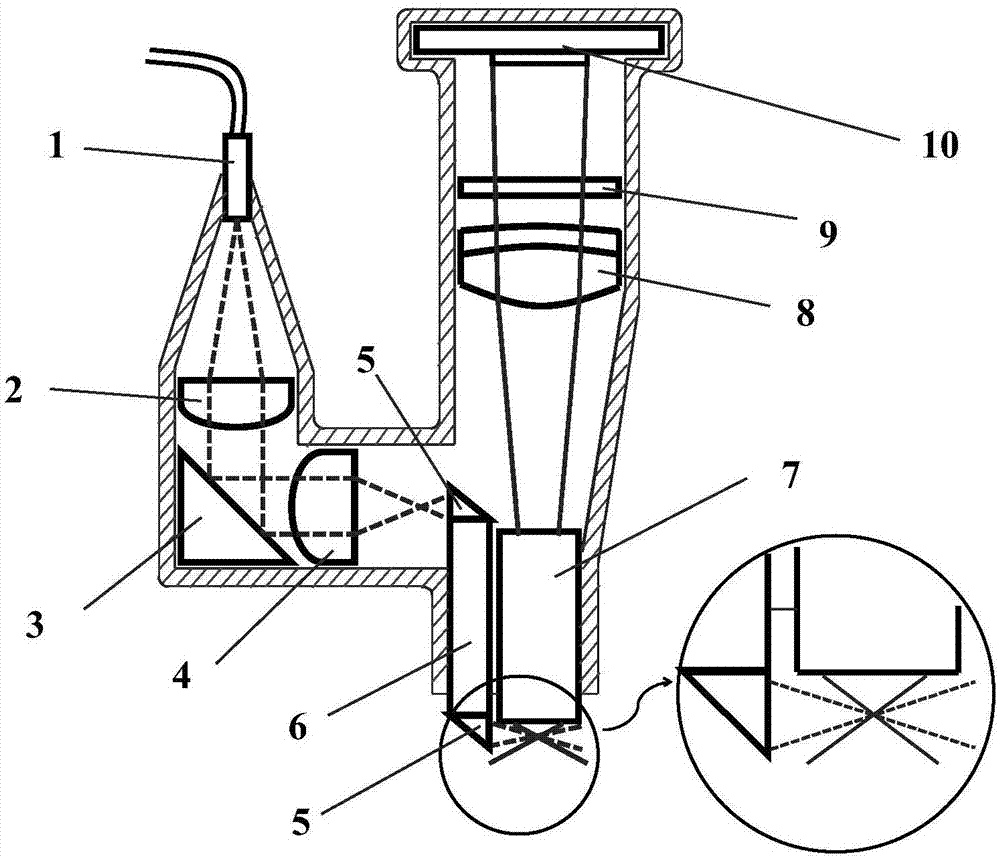
[0049] A head-mounted miniature light sheet microscope provided by the present invention excites the object to be measured by emitting a light sheet from a probe, and collects the fluorescent signal generated by the object to be measured in response to the emitted light sheet signal to obtain the fluorescence imaging information of the object to be measured , the probe is set as a micro-light sheet microscopic imaging probe, and the structural diagram is as follows figure 1 shown.

[0050] The miniature light sheet microscopic imaging probe is used to install on the head of the object to be measured. The laser light is transmitted to the microscopic light sheet microscopic imaging probe through the optical fiber 1. The fluorescent signal is collected in the direction for imaging, and the neurofluorescence image of the object to be tested is obtained.

[0051] The interior of the probe is provided with an illumination optical path unit and an imaging optical path unit; the ill...

Embodiment 2

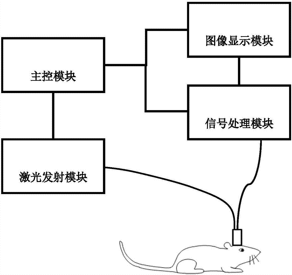
[0058] A kind of miniature light sheet microscope provided in this embodiment, other characteristics are identical with embodiment 1, difference is: also be provided with laser emission module, signal processing module, image display module and main control module, such as figure 2 shown.

[0059] The main control module is electrically connected to the laser emitting module, the image display module, and the signal processing module respectively, and the laser emitting module is connected to the micro-optic microprobe through an optical fiber 1, and the micro-optic A chip microprobe is connected to the signal processing module, and the signal processing module is connected to the image display module;

[0060] The laser emitting module is used to generate and output laser light;

[0061] The micro-light-sheet microscopic imaging probe is used to excite, collect and convert fluorescent signals;

[0062] The main control module is used to control the laser emitting module, t...

Embodiment 3

[0066] A kind of miniature light sheet microscope provided by this embodiment, other features are the same as embodiment 2, the difference is that: the laser emission module is provided with a laser for generating laser, the main control module is set as a computer, and the image display module is set as a display screen .

[0067] The miniature light sheet microscope provided in this embodiment uses fluorescent calcium imaging technology, and uses light sheet scanning to replace the slow point scanning imaging method. When the object to be detected is excited, the light sheet has good optical layer selection ability and large Large excitation area, improved imaging speed and signal-to-noise ratio, reduced phototoxicity, suitable for long-term observation of neural activity in vivo.


PUM


Abstract

Provided is a head-mounted micro light sheet microscope. A probe emits a light sheet to excite an object to be measured, and collects a fluorescence signal produced by the object to be measured according to an emitted light sheet signal to obtain the fluorescence imaging information of the object to be measured. The imaging probe can be fixed to the head of a free moving animal. The object to be measured is excited by the light sheet, and fluorescence is collected in a direction perpendicular to the light sheet to carry out imaging. Light sheet scanning is used to replace the point scanning imaging mode featuring slow imaging. During excitation of the object to be measured, the light sheet has good optical layer selection capability and large excitation area. Therefore, the imaging speed and the signal-to-noise ratio are improved, and the light toxicity is reduced. The head-mounted micro light sheet microscope is suitable for long-time living imaging. A high-contrast image can be obtained. Real-time imaging of the brain of a free moving animal is realized.

Description

technical field [0001] The invention relates to the field of fluorescence imaging, in particular to a head-mounted miniature light sheet microscope. Background technique [0002] Imaging living brain tissue is an important means of studying animal behavior and neural activity in today's brain neuroscience. At present, some miniature fluorescent microscopes used for free-moving animals are small in size and light in weight, and can be fixed on the head of the animal without affecting the freedom of the animal. Activity, which can perform real-time imaging of living organisms. However, the existing miniature fluorescent microscopes that adopt the point-scanning imaging method reduce the imaging speed, have high phototoxicity, and are not suitable for long-term in vivo imaging, and the use of wide-field detection will reduce the ability of layer selection, and the imaging will be affected by the background signal. interference, thereby reducing imaging contrast and signal-to-n...

Claims


Application Information

Patent Timeline
Patent Type & Authority Applications(China)
IPC IPC(8): G02B21/00G02B21/06G02B21/36
Inventor 戚力黄诗娴冯前进陈武凡
Owner SOUTHERN MEDICAL UNIVERSITY
Why Eureka
Try Eureka