A method and device for automatic detection of thin fibrous cap plaques based on cardiovascular OCT images

A cardiovascular and fiber technology, applied in the field of automatic detection of thin fibrous cap plaques based on cardiovascular OCT images, can solve the problems of high cost, inability to establish a unified clinical standard, and inability to meet real-time analysis, achieving good robustness and High detection speed and high detection accuracy

Active Publication Date: 2021-02-02
GENERAL HOSPITAL OF PLA
View PDF4 Cites 0 Cited by
  • Summary
  • Abstract
  • Description
  • Claims
  • Application Information

AI Technical Summary

Problems solved by technology

[0004] Although there are many methods for the detection of thin fibrous cap plaques, including coronary angiography, intravascular ultrasound, optical coherence tomography, etc., they all need to be based on manual identification of whether the image contains thin fibrous cap plaques, and the current clinical evaluation and analysis of cardiac Vascular thin fibrous cap plaques require doctors to spend a lot of time and energy to analyze and judge whether OCT images contain thin fibrous cap plaques; on the other hand, it cannot meet the needs of real-time clinical analysis and cannot establish a unified clinical standard

Method used


Image

  • A method and device for automatic detection of thin fibrous cap plaques based on cardiovascular OCT images
  • A method and device for automatic detection of thin fibrous cap plaques based on cardiovascular OCT images
  • A method and device for automatic detection of thin fibrous cap plaques based on cardiovascular OCT images

Examples


Embodiment 1

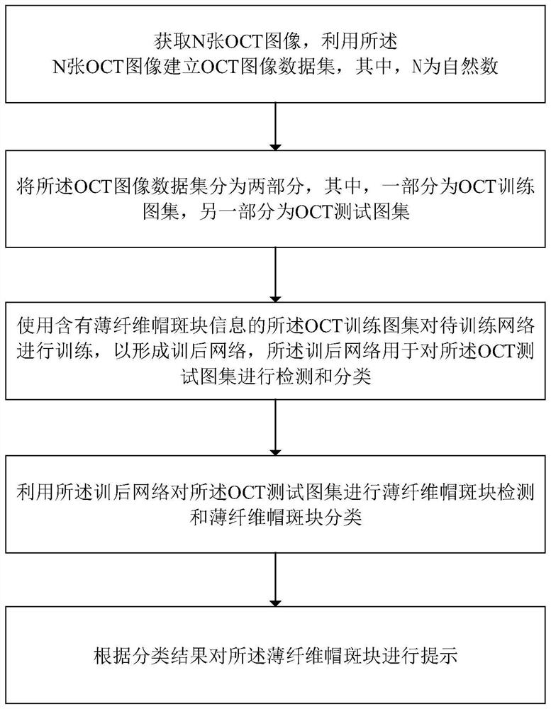
[0058]Seefigure 1 ,figure 1 It is a schematic flowchart of an automatic detection method for thin fiber cap plaque based on cardiovascular OCT images provided by an embodiment of the present invention. The detection method includes:

[0059]Step 1. Obtain N OCT images, and use N OCT images to build an OCT image data set, where N is a natural number;

[0060]Step 2. Divide the OCT image data set into two parts, one of which is the OCT training atlas and the other is the OCT test atlas;

[0061]Step 3. Use the OCT training atlas containing thin fiber cap patch information to train the network to be trained to form a post-training network, which is used to detect and classify the OCT test atlas;

[0062]Step 4. Use the post-training network to detect and classify thin fiber cap plaques on the OCT test atlas;

[0063]Step 5. Prompt the thin fiber cap plaque according to the classification result.

[0064]Preferably, both the OCT training atlas and the OCT test atlas include OCT images with thin fiber cap...

Embodiment 2

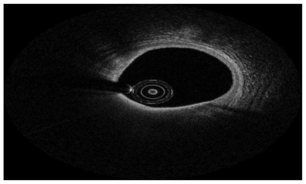
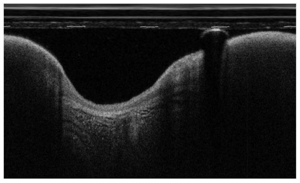
[0088]SeeFigure 2~Figure 14 ,figure 2 Is a schematic diagram of an OCT image in a rectangular coordinate system provided by an embodiment of the present invention,image 3 Is a schematic diagram of an OCT image in a polar coordinate system provided by an embodiment of the present invention,Figure 4 This is a feature map sub-region average pooling provided by an embodiment of the present invention,Figure 5 Is a schematic diagram of a dimensionality change provided by an embodiment of the present invention,Image 6 Is a schematic diagram of the composition of a loss function provided by an embodiment of the present invention,Figure 7 Is a schematic diagram of an original image in an OCT test atlas provided by an embodiment of the present invention,Figure 8 Is a schematic diagram of an OCT image to be detected in an OCT test atlas provided by an embodiment of the present invention,Picture 9 Is a schematic diagram of the original image in another OCT test atlas provided by an embodiment o...

Embodiment 3

[0161]SeeFigure 15 ,Figure 15 It is a schematic flowchart of another method for automatically detecting thin fibrous cap plaque based on cardiovascular OCT images according to an embodiment of the present invention. The detection method includes:

[0162]Step 1. Obtain multiple OCT images and establish an OCT image data set;

[0163]Step 2. Divide the OCT image data set into OCT training atlas and OCT test atlas;

[0164]Step 3. Use the OCT training atlas to train the network to be trained to form a post-training network;

[0165]Step 4. Use the post-training network to detect and classify thin fiber cap plaques on the OCT test atlas;

[0166]Step 5. Prompt the thin fiber cap plaque according to the classification result.

[0167]Among them, after step 1, it also includes:

[0168]Step 1.1: Perform coordinate conversion on the images in the OCT image data set, so as to convert the OCT image data set in the rectangular coordinate system into the OCT image data set in polar coordinates.

[0169]Among them, a...


PUM


Abstract

The present invention relates to a method and device for automatic detection of thin fibrous cap plaques based on cardiovascular OCT images. The method includes: using N OCT images to establish an OCT image data set; Test atlas; use the OCT training atlas containing thin fiber cap plaque information to train the network to be trained to form a post-training network, which is used to detect and classify the OCT test atlas; use the post-training network to detect and classify the OCT test atlas Thin fibrous cap plaque detection and thin fibrous cap plaque classification are performed on the test atlas; thin fibrous cap plaques are prompted according to the classification results. The present invention uses the trained network to extract features from the image, realizes automatic detection and identification of whether there is a thin fibrous cap plaque on the OCT image, does not require manual participation, and is convenient for doctors to accurately analyze the OCT image, and the detection accuracy is relatively high. High, with better robustness and detection speed.

Description

Technical field[0001]The present invention relates to the technical field of medical devices, in particular to a method and device for automatically detecting thin fiber cap plaques based on cardiovascular OCT images.Background technique[0002]Vulnerable plaques refer to all plaques that easily lead to thrombosis or can quickly develop into disease. The envelope on the surface of vulnerable plaque is very thin, and the plaque contains a lot of lipids, so it is easy to rupture. For example, emotional stimulation, strenuous exercise, alcoholism, cold and other factors can cause the human body to increase blood pressure, blood flow violently impacts the plaque, or vasospasm. In these cases, the capsule of the vulnerable plaque will be damaged and ruptured.[0003]At present, thin fibrous cap plaque (TCFA) with inflammation infiltration is the most common type of vulnerable plaque. Acute coronary events caused by its secondary plaque rupture account for 60-70% of all coronary events.[0004]...

Claims


Application Information

Patent Timeline
Patent Type & Authority Patents(China)
IPC IPC(8): G06K9/00G06K9/32G06K9/62
CPCG06V20/698G06V10/25G06V2201/03G06F18/214
Inventor 陈韵岱曹一挥朱锐李嘉男金琴花荆晶
Owner GENERAL HOSPITAL OF PLA
Why Eureka
Try Eureka