Three-dimensional ultrasound reconstruction method and device, equipment and storage medium

A technology of three-dimensional ultrasound and ultrasound images, applied in the field of medical images, can solve the problems of poor quality of three-dimensional ultrasound image reconstruction, not considering the influence of limited ultrasound data, etc. Effect

Active Publication Date: 2018-06-22
SHENZHEN INST OF ADVANCED TECH CHINESE ACAD OF SCI
View PDF5 Cites 8 Cited by
  • Summary
  • Abstract
  • Description
  • Claims
  • Application Information

AI Technical Summary

Problems solved by technology

[0004] The object of the present invention is to provide a three-dimensional ultrasonic reconstruction method, device, image processing equipment and storage medium, aiming to solve the problem that the three-dimensional ultrasonic reconstruction of the prior art does not take into account the limitations of single-angle scanning and the impact of motion artifacts on ultrasonic data. The influence of the influence, resulting in the problem of poor reconstruction quality of 3D ultrasound images

Method used


Image

  • Three-dimensional ultrasound reconstruction method and device, equipment and storage medium
  • Three-dimensional ultrasound reconstruction method and device, equipment and storage medium
  • Three-dimensional ultrasound reconstruction method and device, equipment and storage medium

Examples


Embodiment 1

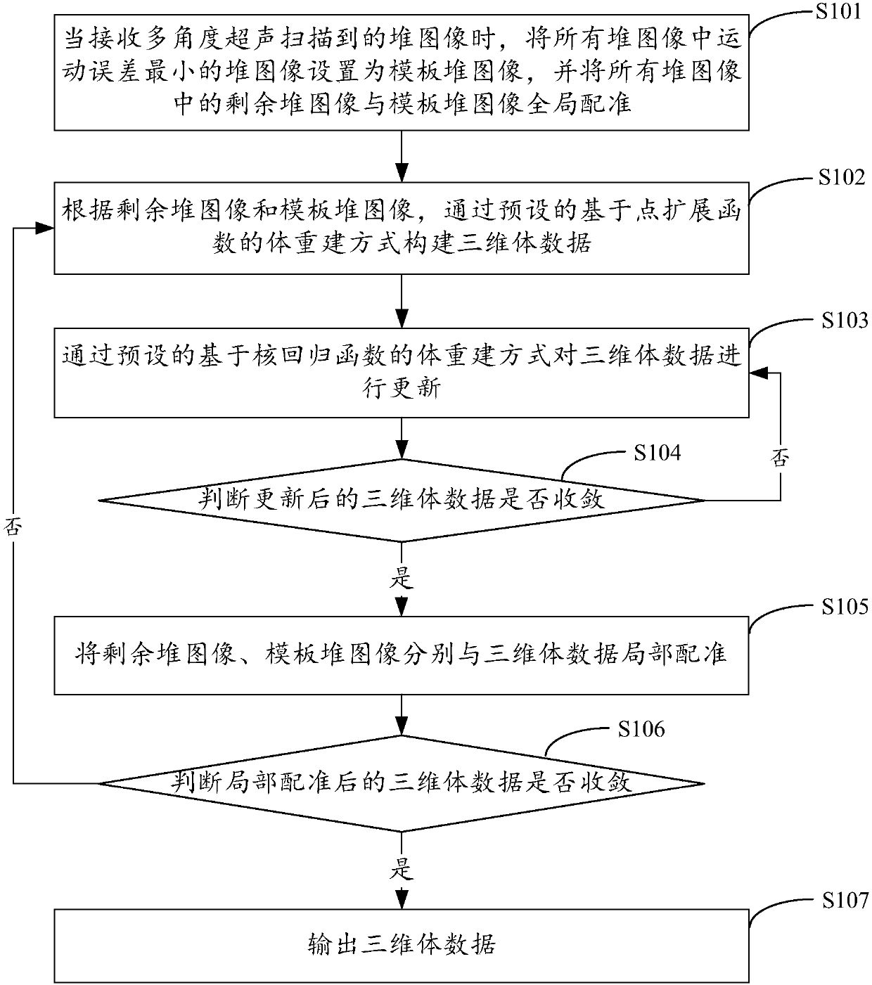
[0027] figure 1 It shows the implementation process of a three-dimensional ultrasonic reconstruction method provided by Embodiment 1 of the present invention. For the convenience of description, only the parts related to the embodiment of the present invention are shown, and the details are as follows:

[0028] In step S101, when receiving stack images scanned by multi-angle ultrasound, set the stack image with the smallest motion error among all stack images as the template stack image, and globally align the remaining stack images in all stack images with the template stack image allow.

[0029] The embodiment of the present invention is applicable to a three-dimensional ultrasound system. Ultrasonic scanning is performed at different angles to obtain a sequence of two-dimensional ultrasonic images scanned at different angles (ie, a sequence of two-dimensional slices). The sequence of two-dimensional ultrasonic images at the same angle can be called a stack image. A method...

Embodiment 2

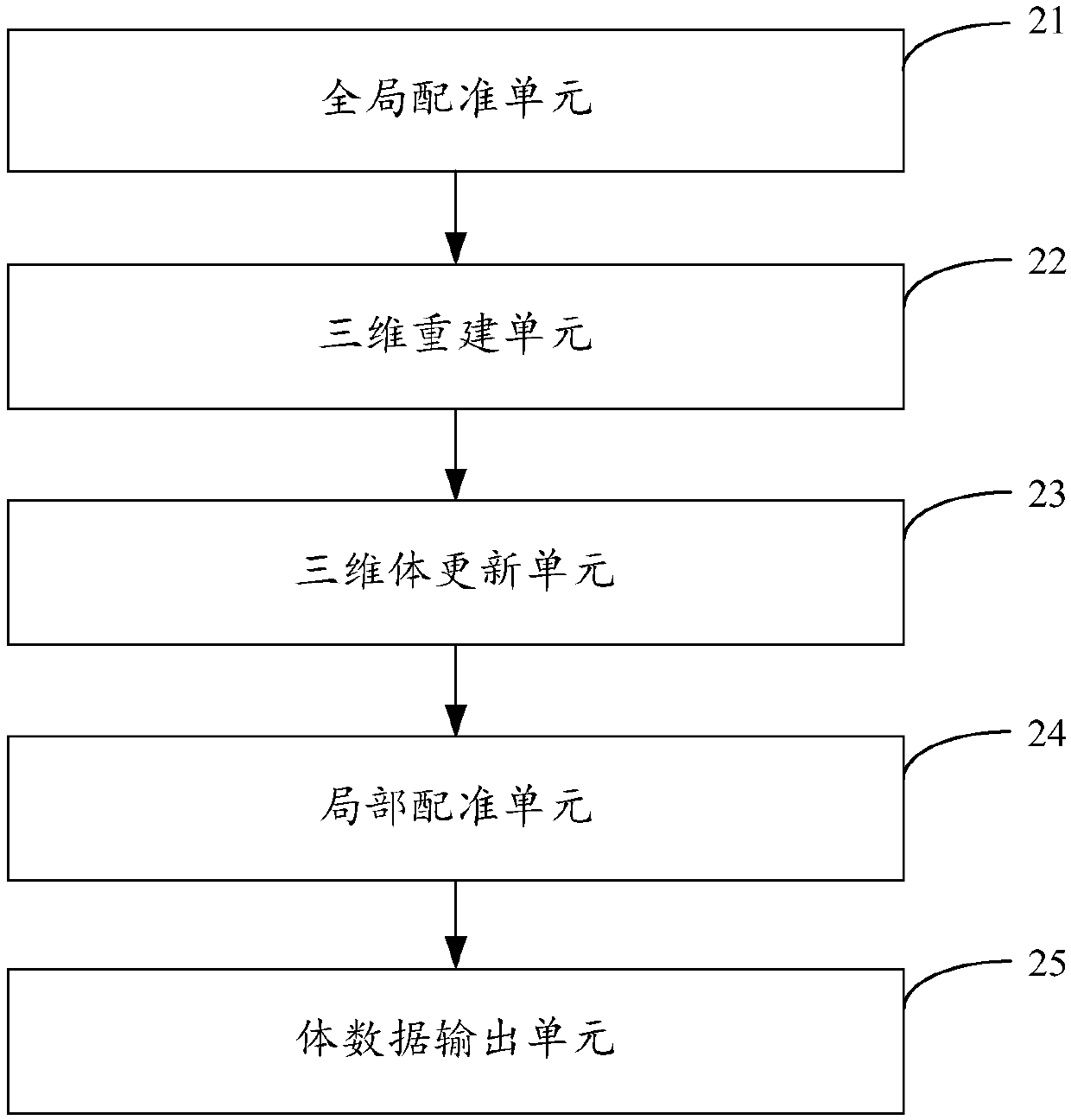
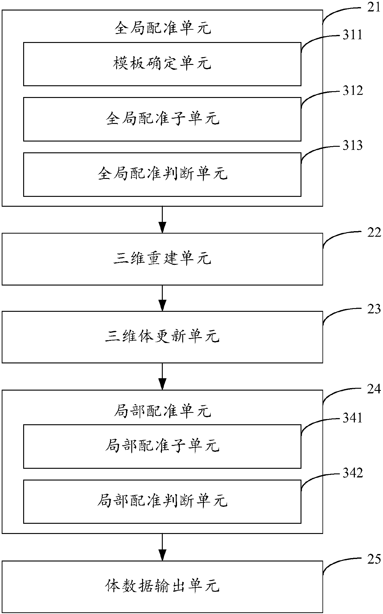
[0067] figure 2 The structure of a three-dimensional ultrasonic reconstruction device provided by Embodiment 2 of the present invention is shown. For the convenience of description, only the parts related to the embodiment of the present invention are shown, including:

[0068] The global registration unit 21 is configured to set the stack image with the smallest motion error among all the stack images as the template stack image when receiving the stack images scanned by the multi-angle ultrasound, and align the remaining stack images among all the stack images with the template stack Image global registration.

[0069] In the embodiment of the present invention, ultrasonic scanning is performed at different angles to obtain stack images scanned at different angles. A method based on motion estimation can be used to select a stack image with the smallest motion error among all stack images as a template stack image, and then globally register the remaining stack images in a...

Embodiment 3

[0114] Figure 4 The structure of the image processing device provided by the third embodiment of the present invention is shown, and for the convenience of description, only the parts related to the embodiment of the present invention are shown.

[0115] The image processing device 4 of the embodiment of the present invention includes a processor 40 , a memory 41 and a computer program 42 stored in the memory 41 and operable on the processor 40 . When the processor 40 executes the computer program 42, the steps in the above-mentioned method embodiments are realized, for example figure 1 Steps S101 to S107 are shown. Alternatively, when the processor 40 executes the computer program 42, the functions of the units in the above-mentioned device embodiments are realized, for example figure 2 Function of units 21 to 25 shown.

[0116] In the embodiment of the present invention, among the stack images scanned by multi-angle ultrasound, the stack image with the smallest motion e...


PUM


Abstract

The invention provides a three-dimensional ultrasound reconstruction method and device, equipment and a storage medium, which are suitable for use in the technical field of computers. The method comprises: setting a heap image of a smallest motion error in heap images, which are scanned by multi-angle ultrasound, as a template heap image, and carrying out global registration on the remaining heapimages with the template heap image; constructing three-dimensional body data according to the remaining heap images and the template heap image; updating the three-dimensional body data through a kernel reconstruction function-based body reconstruction manner, and judging whether the three-dimensional body data after updating converge; if yes, carrying out local registration on the remaining heapimages and the template heap image with the three-dimensional body data, and otherwise, jumping to the step of updating the three-dimensional body data; and judging whether the three-dimensional bodydata after local registration converge, if yes, outputting the three-dimensional body data after local registration, and otherwise, jumping to the step of constructing three-dimensional body data. Therefore, three-dimensional ultrasound reconstruction based on multi-angle scanning is realized, and reconstruction quality of a three-dimensional ultrasound image is effectively improved.

Description

technical field [0001] The invention belongs to the technical field of medical images, and in particular relates to a three-dimensional ultrasonic reconstruction method, device, equipment and storage medium. Background technique [0002] The three-dimensional ultrasound system can intuitively provide doctors with the spatial position and three-dimensional shape of organs or tissues of interest in the body, and more accurately reflect the shape, volume, contour, and adjacency between organs in clinical practice. At present, three-dimensional ultrasound systems mainly include two categories. One is the volumetric imaging method based on a two-dimensional area array probe (also known as a volumetric probe dedicated to three-dimensional ultrasound). The device drives the chip to do equidistant sector scan or circular scan. The other is to use traditional two-dimensional ultrasound equipment combined with specific spatial positioning information to obtain a sequence of two-dimens...

Claims


Application Information

Patent Timeline
Patent Type & Authority Applications(China)
IPC IPC(8): G06T15/00G06T7/30
CPCG06T7/30G06T15/005G06T2207/10012
Inventor 温铁祥刘蓉李凌秦文健
Owner SHENZHEN INST OF ADVANCED TECH CHINESE ACAD OF SCI
Why Eureka
Try Eureka