Dual-frequency intravascular ultrasound imaging probe

An ultrasonic imaging, dual-frequency technology, applied in ultrasonic/sonic/infrasonic diagnosis, catheter, sonic diagnosis and other directions, can solve the problems of low frequency, the detection of atherosclerotic plaques in the early stage of small tissue lesions of the blood vessel wall cannot achieve accurate detection and other problems

Active Publication Date: 2018-07-13
SHENZHEN INST OF ADVANCED TECH
View PDF5 Cites 15 Cited by
  • Summary
  • Abstract
  • Description
  • Claims
  • Application Information

AI Technical Summary

Problems solved by technology

The frequency of the traditional electronic phased array catheter transducer is generally around 20MHz, and the frequency of the mechanical catheter transducer is generally aro

Method used


Image

  • Dual-frequency intravascular ultrasound imaging probe
  • Dual-frequency intravascular ultrasound imaging probe
  • Dual-frequency intravascular ultrasound imaging probe

Examples


Embodiment Construction

[0029] Specific embodiments of the present invention will be described below in conjunction with the accompanying drawings. In the specific embodiments of the present invention described below, some very specific technical features are described in order to better understand the present invention, but it is obvious that not all of them are All technical features are necessary technical features for realizing the present invention. Some specific embodiments of the present invention described below are only some exemplary specific embodiments of the present invention, which should not be regarded as limiting the present invention. Additionally, well-known techniques have not been described in order to avoid obscuring the present invention.

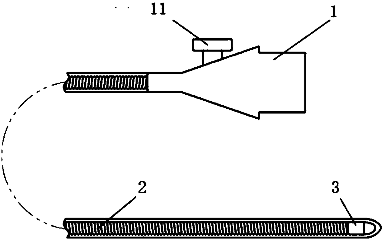
[0030] figure 1 It is a structural schematic diagram of an ultrasonic imaging device with a dual-frequency intravascular ultrasonic imaging probe of the present invention. Such as figure 1 As shown, the ultrasonic imaging device includes...


PUM

PropertyMeasurementUnit
Diameteraaaaaaaaaa

Abstract

The invention provides a dual-frequency intravascular ultrasound imaging probe. The probe comprises a low-frequency ultrasound transducer and a high-frequency ultrasound transducer for simultaneouslyor separately transmitting ultrasound to conduct imaging, and the two ultrasound transducers are connected through the same coaxial cable. According to the probe, a low-frequency single-array-elementtransducer and a high-frequency single-array-element transducer are adopted, excitation signals can be simultaneously or separately transmitted to the two transducers when the probe is operated, so that the transducers perform ultrasound transmission or receiving, and low-frequency and high-frequency ultrasound images can be obtained simultaneously by subsequently conducting processing such as filtering and the like on signals received by the transducers. In addition, two transducers are adopted while only one coaxial cable is adopted, compared with a single-array-element mechanical guiding tube, the size of the probe is not increased, and only a rotary withdrawal device and a signal port are needed at the tail part of the guiding tube.

Description

technical field [0001] The present invention generally relates to the field of medical ultrasound imaging, in particular to a dual-frequency intravascular ultrasound imaging probe. Background technique [0002] Atherosclerosis is a cardiovascular disease with a high mortality rate, and it has a remarkable feature that it is not easy to be diagnosed before the onset. Atherosclerotic plaque rupture has been demonstrated as the underlying pathophysiology in more than 75% of acute coronary syndromes, thus detecting and characterizing rupture-prone plaques is one of the most active areas of research in cardiology and biomedical imaging one. [0003] At present, a variety of medical imaging techniques can be used to diagnose vascular atherosclerotic lesions. Angiography is the main means of detecting vascular atherosclerotic plaque today, and it is used to determine the location and degree of atherosclerotic vascular stenosis. It injects a contrast agent into blood vessels quic...

Claims


Application Information

Patent Timeline
IPC IPC(8): A61B8/12
CPCA61B8/12A61B8/4444A61B8/4477A61B8/4483
Inventor 邱维宝苏敏洪杰韩张利宁郑海荣
Owner SHENZHEN INST OF ADVANCED TECH
Why Eureka
Try Eureka