
An anatomical model for neuroendoscopic surgery

An anatomical model and endoscopic technology, applied in the field of anatomical models for neuroendoscopic surgery, can solve the problems of non-repeatable operation, toxic immersion solution, and high price, and achieve the effect of easy access, low price, and reduced risk

Active Publication Date: 2021-08-31
MEDPRIN REGENERATIVE MEDICAL TECH
View PDF0 Cites 0 Cited by
  • Summary
  • Abstract
  • Description
  • Claims
  • Application Information

AI Technical Summary

Problems solved by technology

Among them, the cadaver head rehearsal is used in the training of neuroendoscopic surgery in the past, but the cadaver head has the following disadvantages: it is expensive, not easy to obtain, cannot be repeated, and the soaking solution is toxic.

Method used


Image

  • An anatomical model for neuroendoscopic surgery
  • An anatomical model for neuroendoscopic surgery

Examples


Embodiment 1

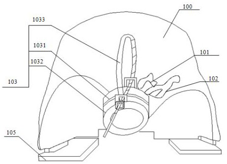
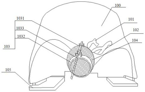
[0037] An anatomical model for neuroendoscopic surgery, including a simulated anterior skull base bone 100, a simulated turbinate, a simulated nasal mucosa, a simulated carotid artery 101, a simulated optic nerve 102, a tumor placement device 103 and a simulated tumor 104, simulating Nasal concha, simulated carotid artery 101, simulated optic nerve 102 are connected with simulated anterior skull base bone 100, tumor placement device 103 is embedded and connected with sella region of simulated anterior skull base bone 100, and simulated nasal mucosa is covered on the simulated turbinate outer wall , the simulated tumor body 104 is placed in the tumor body placement device 103 . The model also includes a simulated face, and the simulated face is fitted and connected with the simulated anterior cranial base bone.

[0038] The preparation method of the anatomical model includes obtaining image data (CT or MRI) of the anterior skull base bone, turbinate, carotid artery, and optic n...

Embodiment 2

[0050] An anatomical model for neuroendoscopic surgery, in addition to the structural composition of Embodiment 1, it also includes angle adjustment plates 105, which are arranged on the left and right sides of the simulated anterior skull base bone 100 bottom.

[0051] The preparation method of the model is the same as that in Example 1, wherein the preparation method of the tumor body is as follows: the egg is boiled until half-cooked, which is used as a simulated tumor body. Among them, the eggshell can simulate the bony structure of the sellar floor (thin bone), the protein can simulate the pituitary tumor, the egg yolk can simulate the pituitary gland, and the egg membrane can simulate the dura mater of the sellar floor. Most sellar tumors, such as pituitary tumors, are soft in texture, similar to half-cooked proteins.

[0052] During the operation, the adjustment plate can adjust the bone quality of the anterior skull base, tilt the head back, and make the nasal passage ...


PUM


Abstract

The invention discloses an anatomical model used for neuroendoscopic surgery. The anatomical model for neuroendoscopic surgery includes simulated anterior skull base bone, simulated turbinate, simulated nasal mucosa, simulated carotid artery, simulated optic nerve, and a tumor placement device for placing simulated tumors. The simulated turbinate, simulated carotid artery, and simulated optic nerve are connected to the simulated anterior skull base bone, the tumor placement device is set at the sella region of the simulated anterior skull base bone, and the simulated nasal mucosa covers the On the outer wall of the simulated turbinate and can be peeled off from the simulated turbinate. The anatomical model prepared by the 3D printing technology of the present invention has good authenticity, is cheap, easy to obtain and has a complete lesion structure, and can be used for long-term and repeated training and operation training of neuroendoscopic simulation surgery, and can be used for Neuroendoscopic surgery training, surgical drills and preoperative simulation operations reduce the risk of surgery.

Description

technical field [0001] The invention relates to the technical field of neurosurgical medical instruments, in particular to an anatomical model for neuroendoscopic surgery. Background technique [0002] Neuroendoscopic surgery is one of the main minimally invasive techniques in neurosurgery, which has the advantages of small trauma, good curative effect, and few postoperative complications. Compared with microscopes, endoscopic surgery has less trauma, clearer field of vision, and wider range of exposure. It can observe lesions at close range, and the surgical operation is precise and meticulous, which greatly reduces surgical injuries, reduces complications, and improves surgical efficacy. Moreover, the postoperative pain is small, the recovery is quick, the hospital stay is short, and the cost is low. Neuroendoscopy has great advantages in operations such as skull base tumors, intraventricular tumors, hydrocephalus, and cerebral hemorrhage, greatly reducing surgical trauma...

Claims


Application Information

Patent Timeline
Patent Type & Authority Patents(China)
IPC IPC(8): G09B23/32G09B23/28
CPCG09B23/285G09B23/32
Inventor 范小敏邓坤学袁玉宇
Owner MEDPRIN REGENERATIVE MEDICAL TECH
Learn More