
Cell image processing method and apparatus

A cell and image technology, applied in character and pattern recognition, instruments, biological neural network models, etc., can solve problems such as high cell overlap rate, weak cytoplasmic edge information, and difficulty in distinguishing cell shapes

Inactive Publication Date: 2018-07-27
BEIHANG UNIV
View PDF4 Cites 19 Cited by
  • Summary
  • Abstract
  • Description
  • Claims
  • Application Information

AI Technical Summary

Problems solved by technology

At present, the challenges of cytoplasmic segmentation mainly lie in: cytoplasm has weak edge information, sometimes it is difficult to distinguish the shape of cells with the naked eye, the overlapping rate of cells is high, and some cells are even completely covered, and various impurities such as blood, inflammatory cells, and human factors interfere

Method used


Image

  • Cell image processing method and apparatus
  • Cell image processing method and apparatus
  • Cell image processing method and apparatus

Examples


Embodiment Construction

[0025]In order to make the purpose, technical solution and advantages of the present invention clearer, the specific implementation of the cell image processing method and device of the present invention will be further described in detail through the following examples and in conjunction with the accompanying drawings. It should be understood that the specific embodiments described here are only used to explain the present invention, not to limit the present invention.

[0026] The present invention relates to the technical fields of cytology, computer vision and image processing, and artificial intelligence. Specifically, it relates to a method and device for processing cell images. More specifically, it is a method and device for automatic segmentation based on overlapping cervical cell images. .

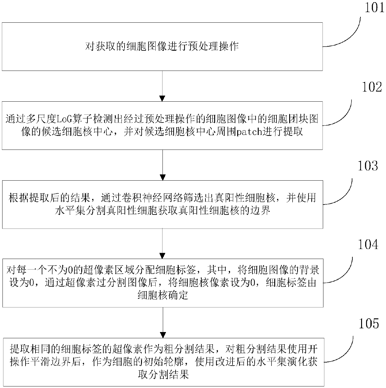
[0027] Such as figure 1 Shown is a schematic flowchart of a cell image processing method in an embodiment. Specifically include the following steps:

[0028] Step 101, preproc...


PUM


Abstract

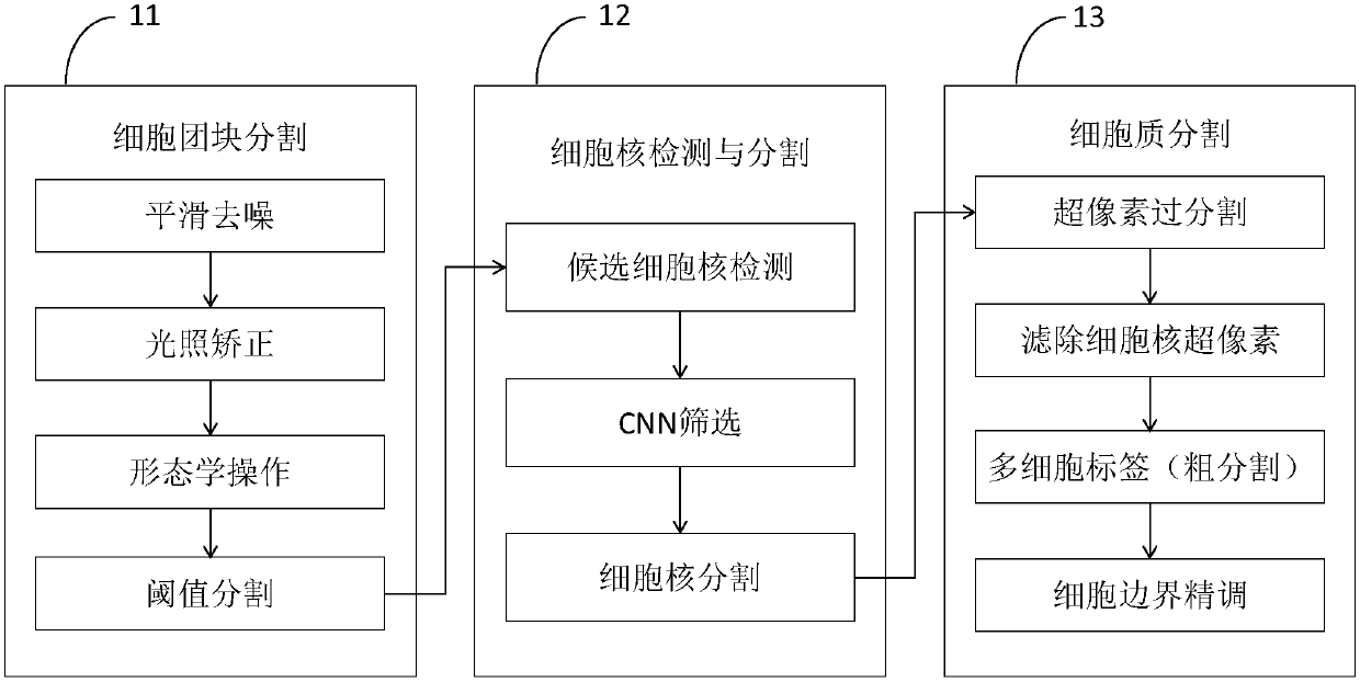
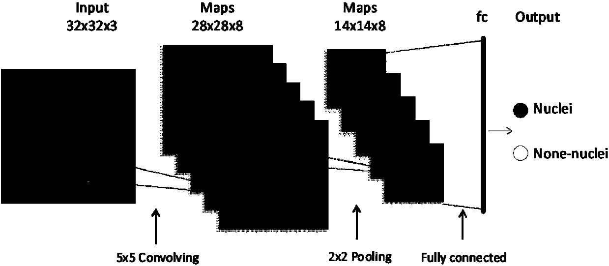
The invention discloses a cell image processing method. The method comprises the steps of preprocessing an image, and separating a cell mass from a background by using an Ostu threshold method and a morphological operation; detecting out candidate cell nucleus centers by using a multi-scale LoG operator, extracting patches around the nucleus centers, screening out true positive cell nucleuses through a convolutional neural network, and segmenting true positive cells by using a level set to obtain boundaries of the cell nucleuses; for segmentation of overlapped cytoplasms, separating all cellsbased on a superpixel multi-cell tag segmentation method, allocating cell tags to all superpixel regions after superpixel image over-segmentation, taking the regions of the same tags as initial contours of the cells, and obtaining more accurate cell boundaries by using improved level set evolution. According to the method provided by the invention, the segmentation problem is converted into the classification problem; the problem due to weak cytoplasm edges is effectively solved; and the segmentation accuracy is improved. The method is high in running speed and has relatively good robustness.The invention furthermore discloses a cell image processing apparatus.

Description

technical field [0001] The invention relates to the technical field of computer image processing, in particular to a cell image processing method and device. Background technique [0002] Worldwide, cervical cancer is the fourth most common cancer and the fourth most common cause of cancer death in women. In 2012, there were approximately 528,000 cases of cervical cancer and 266,000 deaths, accounting for 8% of cancer incidence and cause of death, respectively. About 70% of cervical cancers occur in developing countries and are the most common cause of cancer death in low-income countries. In developed countries, because of the popularity of cervical smear screening, the incidence of cervical cancer has been greatly reduced. [0003] Cervical cancer, also known as cervical cancer (Cervical cancer), is cancer that occurs in the cervix of women. Derived from the growth of abnormal cells, which can even invade or metastasize to other parts of the body. There are usually no ...

Claims


Application Information

Patent Timeline
IPC IPC(8): G06K9/00G06N3/04
CPCG06V20/695G06V20/698G06N3/045
Inventor 万涛秦曾昌徐淑淞
Owner BEIHANG UNIV
Learn More