
Abdomen CT (Computed Tomography) image multi-organ segmentation method based on superpixel

A superpixel segmentation and CT image technology, applied in image analysis, image enhancement, image data processing, etc., can solve problems such as single organs, achieve accurate results, avoid difficult segmentation, and reduce the amount of calculation

Active Publication Date: 2018-08-03
NORTHWEST UNIV
View PDF2 Cites 23 Cited by
  • Summary
  • Abstract
  • Description
  • Claims
  • Application Information

AI Technical Summary

Problems solved by technology

[0005] The object of the present invention is to provide a superpixel-based abdominal CT multi-organ segmentation method to solve the problem in the prior art that the abdominal organ segmentation is mostly performed on a single organ

Method used


Image

  • Abdomen CT (Computed Tomography) image multi-organ segmentation method based on superpixel
  • Abdomen CT (Computed Tomography) image multi-organ segmentation method based on superpixel
  • Abdomen CT (Computed Tomography) image multi-organ segmentation method based on superpixel

Examples


Embodiment Construction

[0052] Comply with the above technical solutions, such as Figure 1 to Figure 8 As shown, the present invention discloses a method for multi-organ segmentation of abdominal CT images based on superpixels, such as figure 1 shown, including the following steps:

[0053] Step 1, collecting multiple abdominal CT images to obtain an abdominal CT image set;

[0054] A CT image is a collection of images obtained by scanning a certain thickness of a certain part of the human body with an X-ray beam using computerized tomography equipment.

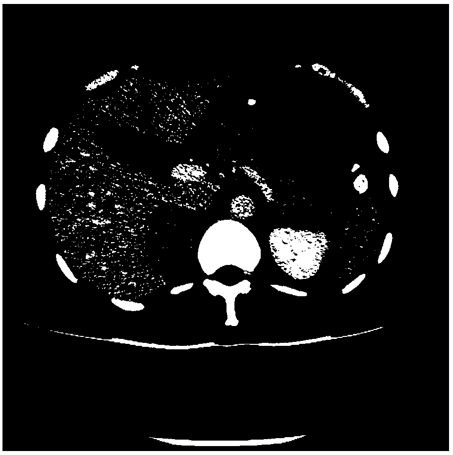
[0055] In this embodiment, a total of 161 abdominal CT images were collected by Toshiba Aquilion64 spiral CT machine, wherein the size of each abdominal CT image was 512×512, and the slice thickness was 1 mm, as figure 2 as shown, figure 2 It is one abdominal CT image in the abdominal CT image set in this embodiment.

[0056] Step 2. Perform preprocessing on each abdominal CT image in the abdominal CT image set to obtain a preprocessed abdomi...


PUM


Abstract

The invention discloses an abdomen CT (Computed Tomography) image multi-organ segmentation method based on a superpixel. The method comprises the following steps that: preprocessing an abdomen CT image, wherein the preprocessing comprises the filtering and the gray scale mapping of the abdomen CT image; utilizing correlation between an upper layer and a lower layer in an image in an abdomen CT image set; in addition, using a K-means clustering method which takes fused Euclidean distance and gray scale distance as a measurement distance to segment the abdomen CT image into superpixel blocks; after the superpixel blocks are subjected to feature extraction, adopting an extreme learning machine to be combined with a position and gray scale statistical probability model to classify the superpixel blocks; and then, combining classified superpixel blocks, and obtaining an abdomen CT image multi-organ segmentation result. By use of the method provided by the invention, the abdomen CT image canbe accurately subjected to multi-organ segmentation, a calculated amount is lowered to a pixel block level from a pixel level, the calculated amount is greatly lowered, and abdomen CT image multi-organ segmentation speed is quickened.

Description

technical field [0001] The invention relates to an image segmentation method, in particular to a multi-organ segmentation method for abdominal CT images based on superpixels. Background technique [0002] With the development of medical imaging technology, medical imaging plays an indispensable role in medical diagnosis. At the same time, the rapid development of computer-aided diagnosis (Computer Aided Diagnosis, CAD) technology can use computer-aided processing and analysis of medical images to help doctors better correct diagnosis of the disease. As the first stage of medical image processing, medical image segmentation is of great significance to medical image analysis and visualization. Medical image segmentation is the basis of lesion area extraction, organ tissue measurement and 3D reconstruction. Standards and other aspects also played an irreplaceable role. [0003] The distribution of human abdominal organs is complex, mainly including liver, kidney, gallbladder,...

Claims


Application Information

Patent Timeline
IPC IPC(8): G06T7/10
CPCG06T2207/10081G06T2207/20028G06T7/10
Inventor 张蕾吕朝晖冯筠卜起荣王红玉
Owner NORTHWEST UNIV
Learn More